Orbital Hemorrhage
Updated May 2024
Michael A. Burnstine, MD, FACS; Priya D. Sahu, MD
Orbital hemorrhage is bleeding within the orbit that can quickly cause vision loss if not addressed in a timely manner. Vision loss occurs due to a compartment syndrome within the orbital walls that compromises the optic nerve and its blood supply. Detailed knowledge of orbital anatomy is necessary to properly diagnose and safely treat an orbital hemorrhage.
Orbital Anatomy
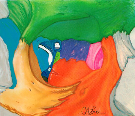
Orbital bones
(Figure 1)
- Frontal
- Zygomatic
- Maxilla
- Ethmoid
- Sphenoid
- Lacrimal
- Palatine

Figure 1. Orbital anatomy.
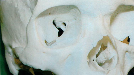
Windows into the orbit
(Figure 2)
Orbital foramina
- Optic foramen
- Begins at middle cranial fossa and terminates at the apex of the orbit
- Through the foramen, the optic nerve, (CN II), the ophthalmic artery, and sympathetic fibers from the carotid plexus travel
- Supraorbital foramen
- Located at the medial third of the superior margin of the orbit
- Transmits the supraorbital nerve, a branch of the ophthalmic division of CN V, and supraorbital artery and vein
- Anterior ethmoidal foramen
- Located at the frontoethmoidal suture
- Transmits the anterior ethmoidal vessels and nerve
- About 12 mm posterior to the anterior orbital rim
- Posterior ethmoidal foramen
- Located at the junction of the roof (frontal bone) and medial wall (ethmoid) of the orbit
- Transmits the posterior ethmoidal vessels and nerve through the frontal bone
- About 6 mm posterior to the anterior ethmoidal foramen
- Zygomatic foramen
- Located in the lateral aspect of the zygomatic bone
- Transmits the zygomaticofacial and zygomaticotemporal branches of the zygomatic nerve and artery

Figure 2. Windows into the orbit.
Ducts
- Nasolacrimal duct
- Located in the inferior lacrimal fossa and travels postero-laterally ending below the inferior turbinate
- Transmits the nasolacrimal duct
Canals
- Infraorbital canal
- Is a continuation of the infraorbital groove and exits about 4 mm below the inferior orbital margin
- Transmits the infraorbital nerve, a branch of the maxillary division of the cranial nerve V
Fissures
- Superior orbital
- Fissure located between the greater and lesser wings of the sphenoid bone; lies below and lateral to the optic foramen. It is 22 mm long and is spanned by the annulus of Zinn
- Transmits cranial nerves and vessels:
- Lacrimal nerve of CN V (trigeminal)
- Frontal nerve of CN V
- CN IV (trochlear)
- Superior Ophthalmic Vein
- Superior and Inferior divisions of CN III (oculomotor)
- Nasociliary branch of CN V
- Sympathetic Roots of Ciliary Ganglion
- CN VI (Abducens)
- Inferior orbital fissure
- Located below the superior orbital fissure between the lateral wall and the floor of the orbit with access to the pterygopalatine and inferotemporal fossa
- Transmits the infraorbital and zygomatic branches of the CN V, (maxillary division), orbital nerve from the pterygopalatine fossa, and the inferior ophthalmic vein
Orbital contents
- Periorbita
- The periosteal covering of the orbital bones that is continuous with the orbital septum at the arcus marginalis. It fuses posteriorly with the dura mater covering the optic nerve
- Connective tissue
- Annulus of Zinn
- Fibrous ring formed by the common origin of the 4 rectus muscles
- Encircles the optic foramen and central portion of the superior orbital fissure
- The oculomotor foramen transmits both upper and lower divisions of CN III, CN VI, and the nasociliary branch of the ophthalmic division of CN V
- Intermuscular septum
- Connects the rectus muscles in the anterior orbit
- Septum divides the orbital fat into the intraconal fat and the extraconal fat
- Orbital septum
- Thin sheet of connective tissue originates from the periosteum of the roof and floor of the orbit along the arcus marginalis of the orbital rim
- Inserts onto the aponeurosis and the anterior surface of the levator muscle
- Posterior to the orbital septum lies orbital fat, extraocular muscles, and a rich blood supply
- Provides a barrier between the anterior and posterior orbit for spread of blood/inflammation
- Extraocular muscles and orbital fat
- Four rectus muscles (superior, inferior, medial, and lateral originate in the annulus of Zinn and insert onto the globe
- The superior oblique muscle originates slightly medial to the levator muscle and travels through the trochlea on the superomedial orbital rim where it turns posterolateral and inserts on the globe
- The inferior oblique originates on the anterior orbital floor lateral to the lacrimal sac and travels posterolaterally within the lower eyelid retractors to insert temporal to the macula
- The levator muscle arises above the annulus on the lesser wing of the sphenoid; its aponeurosis inserts onto the tarsal border of the upper eyelid and the skin
- Nerves within the orbit
- Optic nerve (CN II)
- Sensory nerve
- 30 mm in length
- The dura mater covering the posterior portion of the intraorbital optic nerve fuses with the annulus of Zinn at the orbital apex
- The dura at the apex is continuous with the periosteum of the optic canal
- Oculomotor nerve (CN III)
- Motor nerve
- Superior division innervates superior rectus and levator palpebrae superioris
- Inferior division innervates inferior rectus, medial rectus, and inferior oblique muscles
- Trochlear nerve (CN IV)
- Motor nerve
- Innervates superior oblique muscle
- Trigeminal nerve
- Sensory and motor nerve
- Ophthalmic division
- Frontal nerve innervates medial upper lid and conjunctiva, forehead, scalp, frontal sinuses, and side of nose
- Lacrimal nerve innervates lacrimal gland, adjacent conjunctiva and skin
- Nasociliary nerve carries sensation for the lacrimal drainage system, the conjunctiva, and medial canthal skin
- Short ciliary nerves carry globe sensation
- Long ciliary nerves carry sensation from the ciliary body, iris, and cornea with simultaneous sympathetic innervation to iris dilator muscle
- Maxillary division
- Zygomatic nerve carries sensation of the lateral face
- Maxillary nerve carries sensation of the cheek, lower lid, upper teeth and gums
- Abducens nerve (CN VI)
- Motor nerve
- Innervates the lateral rectus muscle
- Vasculature of the orbit
- The orbit has a rich blood supply.
- Arterial supply
- The blood supply to the orbit is primarily from the ophthalmic artery via the internal carotid artery.
- Major branches of the ophthalmic artery are branches to the extraocular muscles, central retinal artery, and posterior ciliary arteries.
- Terminal branches of the ophthalmic artery form rich anastomoses with branches of the external carotid in the face and periorbital region.
- Contributions also come from the internal maxillary artery and facial artery which are branches of the external carotid artery
- Venous drainage
- The superior and inferior ophthalmic veins provide the main venous drainage of the orbit.
- Both the superior and inferior ophthalmic veins drain into the cavernous sinus.
- Globe
- Sits in the anterior orbit
- Is bound to orbit by optic nerve and its extraocular muscle attachments
Orbital hemorrhage diagnosis
History
- Trauma and mechanism of injury
- Recent eyelid, lacrimal, orbital, or sinus surgery (Rene, BJO 2001)
- Sudden ocular or orbital pain
- Diplopia
- Progressive or acute proptosis
- Nausea/vomiting
Physical exam and testing
- Complete ophthalmic exam. Check the following:
- Decreased visual acuity
- Afferent papillary defect
- Loss of color vision
- Limited extraocular motility
- Eyelid ecchymosis
- Progressive proptosis with resistance to retropulsion
- Chemosis
- Diffuse subconjunctival hemorrhage
- Elevated intraocular pressure
- Optic disc and/or retinal pallor
- Central retinal artery pulsations
- Choroidal folds
- Rule out ruptured globe.
- Scans
- CT scan of orbits to evaluate for retrobulbar hemorrhage (if time allows)
- If vision is threatened, do not wait for orbital CT and perform immediate decompression of compartment syndrome.
- Consider magnetic resonance angiography or magnetic resonance venography of the orbit with spontaneous orbital hemorrhage to look for orbital lymphangiomas, cavernous hemangiomas, and other vascular malformations.
- Lab tests
- CBC with differential
- PT, PTT, platelets
- Differential diagnosis of orbital hemorrhage
- Orbital tumor (Yamamoto, Neurol Med Chir 2012)
- Spontaneous bleed (Elia, Orbit 2013)
- Carotid-cavernous fistula
- Low flow indirect
- High flow direct
- Orbital cellulitis
- Optic nerve sheath hemorrhage
- Terson syndrome
- Retrobulbar injection
Etiology of acute orbital hemorrhage
- Orbital trauma (such as medial wall fracture with laceration of ethmoidal artery)
- Spontaneous bleed following Valsalva from orbital vascular anomaly
- Iatrogenic
- Retrobulbar injections (Kallio, Br J Anesthesia 2000)
- Postsurgical
- Facial fracture repair (Wood, British J Oral and Maxillofac Surg 1989)
- Post-blepharoplasty
- Endoscopic sinus surgery (Rene, BJO 2001)
Risk factors for acute orbital hemorrhage
- Orbital trauma
- Penetrating orbital foreign bodies
- Postseptal eyelid surgery
- Orbital/sinus surgery (Rene, BJO 2001)
- Hypertension
- Anticoagulant medications (Maurer, Int J Oral Maxillofac Surg 2013)
- Aspirin
- NSAIDs
- Warfarin (Coumadin)
- Dabigatran (Pradaxa)
- Rivaroxaban (Xarelto)
- Apixaban (Eliquis)
- Postoperative Valsalva maneuver (vomit, cough, sneeze)
- Coagulopathy (Goyal, Orbit 2004)
- Blood dyscrasia (thrombocytopenia, leukemia) (Grove, Orbit 2008)
- Cirrhosis
Risk factors for spontaneous orbital hemorrhage
- Orbital lymphangiomas with bleed (McNab, Surv Ophthalmol 2014)
- Cavernous hemangiomas (Arora, Orbit 2011)
- Ophthalmic artery aneurysms (McNab, Surv Ophthalmol 2014)
- Hypertension
- Clotting Disorders
- Vitamin-K deficiency
- Anemia
- Hemophilia
- Von Willebrand disease
Patient management
If hemorrhage is limited, not progressive, and does not threaten optic nerve function, close observation can be appropriate.
If vision is threatened and there is progressive tense proptosis, perform urgent canthotomy and cantholysis of the inferior crus of the lateral canthal tendon. This might need to be performed prior to imaging if necessary. If more release is needed, cantholysis of the superior crus of lateral canthal tendon can be performed. If hemorrhage progresses without relief of intraocular pressure, an orbital fracture can be induced for decompression. It is wiser to err on the side of intervention. (Yung, OPRS 1994), (Liu, AJO 1993)
Simultaneously, start medical therapy with acetazolamide 500 mg IV, methylprednisolone 100 mg IV for neuroprotection of the optic nerve, and topical beta-blocker (timolol 0.5% one gtt. q 30 minutes x 2) (Wood, British J Oral and Maxillofac Surg 1989). Consider methylprednisolone 30 mg/kg/q 6 hrs for optic-nerve protection, although benefits are controversial. (Steinsapir, Surv Ophthalmology 1994)
If postoperative bleeding occurred following eyelid, lacrimal, orbital, or sinus surgery, consider orbital exploration to ligate/cauterize source of bleeding.
Consider referral to hematology/oncology to evaluate for blood dyscrasia if spontaneous retrobulbar hemorrhage.
For patients with progressive visual loss, recommend admission for frequent vision checks and consider IV pulse therapy with methylprednisolone 250 mg IV q 6 hrs x 3 day.
Postoperative care
- Steroid/antibiotic eye drops
- IV or PO antibiotics for 1 week
- Ice packs for 48 hours
- If lateral canthal position does not revert to its native position, can perform delayed repair once bleeding has been stabilized
References and additional resources
- AAO, Basic and Clinical Science Course. 2010-11; 7: Orbit, Eyelids, and Lacrimal System.
- AAO, Basic and Clinical Science Course. 2010-2011; 2: Fundamentals and Principles of Ophthalmology.
- Arora V, Prat MD, Kazim M. Acute presentation of cavernous hemangioma of the orbit. Orbit. 2011; 30(4):195-197.
- Brodt J, Gologorsky D, Walter S, Pham S, Gologorsky E. Orbital compartment syndrome following extracorporeal support. J Card Surg. 2013, 28:522-524.
- Elia M, Shield D, Kazim M, Shinder R, Yoon M, McCulley TJ, Shore JW, Greene D, Servat JJ, Levin F. Spontaneous Subperiosteal Orbital Hemorrhage. Orbit 2013; 32(5): 333-35.
- Goyal S, Goel R. Orbital haemorrhage with loss of vision in a patient with disseminated intravascular coagulation and prostatic carcinoma. Orbit. 2004; 23 (3)193-197.
- Grove J, Meyer D. Aplastic anemia presenting as spontaneous orbital hemorrhage. Orbit. 2008; 27 (5):391-393.
- Jamal BT, Diecidue RJ, Taub D, Champion A, Bilyk JR. Orbital hemorrhage and compressive optic neuropathy in patients with midfacial fractures receiving low-molecular weight heparin therapy. J Oral Maxillofac Surg. 2009; 67:1416-1419.
- Kallio, H, Paloheimo M, Maunuksela EL. Hemorrhage and risk factors associated with retrobulbar/peribulbar block: a prospective study in 1383 patients. Br J Anaesth. 2000; 85 (5):708-711.
- Leong JK, Ghabrial R, McCluskey PJ, Mulligan S. Orbital haemorrhage complication following postoperative thrombolysis. Br J Ophthalmology. 2003; 87(5):655-656.
- Liu, D. A simplified technique of orbital decompression for severe retrobulbar hemorrhage. Am J Ophthalmol,1993;116 (1):34.
- Maguire JI, Murchison A P, Jaeger EA. Wills Eye Institute 5-Minute Ophthalmology Consult. Philadelphia: Lippincott, Williams, and Wilkins 2012.
- Maharshak I, Hoang JK, Bhatti MT. Complication of vision loss and ophthalmoplegia during endoscopic sinus surgery. Clin Ophthalmol. 2013; 7;573-580.
- Maurer P, Conrad-Hengerer I, Hollstein S, Mizziani T, Hoffman E, Hengerer F. Orbital hemorrhage associated with orbital fractures in geriatric patients on antiplatelet or anticoagulant therapy. Int J Oral Maxillofac Surg 2013; 42: 1510-1514.
- McNab, A. Nontraumatic orbital hemorrhage. Surv Ophthalmol. 2014; 59:166-184.
- Rene C, Rose GE, Lenthall R, Moseley I. Major orbital complications of endoscopic sinus surgery. Br J Ophthalmol. 2001; 85:598-603.
- Sood V, Rejali D, Stocker J, Pagliarini S, Ahluwalia H, Mehta P. A case report of orbital haemorrhage associated with endoscopic sinus surgery and reversible visual loss: a multidisciplinary approach to management. Orbit. 2013; 32 (1):73-5.
- Steinsapir KD, Goldberg RA. Traumatic optic neuropathy. Surv Ophthalmol. 1994; 38 (6):487-518.
- Wood, CM. The medical management of retrobulbar haemorrhage complicating facial fractures: a case report. Br J Oral Maxillofac Surg. 1989; 27:291-295.
- Yamamoto J, Takahashi M, Nakano Y, Saito T, Kitagawa T, Ueta K, Miyaoka R, Nishizawa S. Spontaneous hemorrhage from orbital cavernous hemangioma resulting in sudden onset of ophthalmopathy in an adult. Neurol Med Chir 2012; 52:741-744.
- Yung CW, Moorthy RS, Lindley D, Ringle M, Nunery WR. Efficacy of lateral canthotomy and cantholysis in orbital hemorrhage. Ophthal Plast Reconstr Surg 1994; 10 (2):137-141.
- Zimmerer R, Schattmann K, Essig H, Jehn P, Metzger M, Kokemuller H, Gellrich N, Tavassol F. Efficacy of Transcutaneous Transseptal Orbital Decompression in Treating Acute Retrobulbar Hemorrhage and Literature Review. Craniomaxillofac Trauma Reconstr. 2014;7:17-26.
