
Endoscopic, Endonasal, or Transnasal Dacryocystorhinostomy (EndoDCR)
Updated August 2026
Robert G. Fante, MD, FACS; Richard C. Allen, MD, Phd
Over the past 25 years, there has been renewed interest in the transnasal approach to dacryocystorhinostomy (DCR) with many variations in surgical technique that are united in their avoidance of an external incision. The use of an endoscope with video magnification can facilitate recognition of surgical landmarks, but many surgeons find that several of the surgical steps are more easily performed with headlight observation. This article reviews the transnasal approach, with or without endoscopic assistance, to treat acquired epiphora related to nasolacrimal duct obstruction.
Goals
Primary:
- Sustained relief from symptomatic epiphora
- Cure for acute or chronic dacryocystitis
- Postsurgical patient satisfaction and relative value has been established by quality of life studies (Jutley, Eye 2013)
Indications and contraindications
Indications are similar to those for external DCR:
- Tearing caused at least in part by partial or complete nasolacrimal duct (NLD) obstruction
- Acute dacryocystitis during or after antibiotic treatment
- NLD obstruction with chronic dacryocystitis
- Tearing without identifiable cause after complete workup (see comments below in Clinical Examination section)
EndoDCR might have advantages over external DCR in some circumstances:
- Younger patients
- Patients with flat nasal bridge
- However, some patients with a wide central midface can have intervening ethmoid air cells between the lacrimal sac and nasal cavity, making endoDCR more challenging
- To avoid disturbing a stable, asymptomatic medial canthal deformity from prior trauma, or surgery such as skin tumor resection/radiation therapy
Relative contraindications:
- Complete canalicular obstruction
- Can be combined with CDCR/Jones tube; see below
- Management of lacrimal sac tumor, lacrimal system stone, or foreign body
- May be more difficult via endonasal route than external DCR, at least for novice surgeons
- In setting of trauma/previous trauma repair when fractures or bone hypertrophy distort anticipated anatomy
- Imaging recommended
- More difficult in setting of nasal septal deviation, unless concurrent septoplasty performed
Preprocedural evaluation
- Complete ophthalmic/medical history with attention to history of
- Topical glaucoma/anti-viral therapy
- Ocular surface disease
- Herpes simplex virus (HSV) infection
- Paranasal sinus disease/surgery
- Facial trauma/surgery/radiation therapy
- Acetylsalicylic acid (ASA)/anticoagulant therapy
- Lacrimal sac tumor
- Granulomatosis with polyangiitis
- Sarcoidosis
- Facial neuropathy
- Allergic fungal sinusitis
- Radioactive iodine therapy
- Clinical examination (see related article, Evaluation of Epiphora)
- Oculofacial external exam
- Slit lamp biomicroscopy of eyelids, puncta, tear film, and anterior segment
- Complete lacrimal evaluation
- Canalicular probing/irrigation
- Jones’ tests
- Nasal examination (office endoscopy preferred)
- Note location of nasal septum, size and configuration of middle turbinate and uncinate process (Figures 1 and 2), presence of polyps or other pathology in the area of the inferior and middle
- DCR studied in patients with epiphora who have a patent lacrimal outflow system to clinical testing and no other cause for epiphora:
- 94% success among 51 patients (O’Donnell, Clin Experiment Ophthalmol 2001)
- Success in these patients has been correlated with the single drop Jones test, especially in the presence of delayed fluorescein dye disappearance and canalicular reflux on irrigation (O’Donnell OPRS 2007)
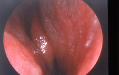
Figure 1. Narrow middle meatus and large turbinate.
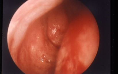
Figure 2. Wide middle meatus and small turbinate with prominent uncinate process.
- Potential imaging options [uncommonly performed in routine cases among ASOPRS members (Nagi, OPRS 2010)]
- Orbit/sinus computed tomography (CT)/magnetic resonance imaging (MRI) scan
- Dacryocystography
- Radionuclide dacryoscintigraphy
Alternatives to procedure
- Observation
- Medical therapy of dacryocystitis, if present
- Lacrimal probing with Mitomycin C
- Standard probing followed by irrigation of 0.5–1 cc mitomycin C 0.2 mg/ml into the nasolacrimal duct, held there with nasal packing for 10 minutes, followed by irrigation of the conjunctival fornices with BSS
- 32/36 eyes (89%) had postoperative patent NLD at 9 months after probing with mitomycin C, but only 9 (25%) were symptom-free (Tsai, Ophthalmology, 2002)
- Balloon dacryoplasty
- Anterograde: 69% improved among 102 cases with at least 12 months follow-up (McCullough, Clin Radiol 2001)
- Retrograde: In a study of 52 cases, 69% failure among complete NLDO and 17% failure for partial NLDO (Fenton, Eye 2001)
- Balloon dacryoplasty plus silicone intubation
- 73% improvement among 30 patients at 1 year (Kuchar, BJO 2001)
- Polyurethane stent (available outside US)
- 55% long-term improvement among 426 patients and recommended by authors for unsuitable surgical candidates (Lanciego, J Vasc Interv Radiol 2003)
- Dacryocystectomy
- External DCR
- Conjunctivodacryocystorhinostomy (CDCR)
- Best alternative when there is severe canalicular obstruction
- Can be performed in conjunction with endoDCR (Trotter, Ophthalmology 2000)
- Botulinum toxin injection into the lacrimal gland
- Munk score improved in 17 patients who underwent botulinum toxin injection into the lacrimal gland rather than surgery for lacrimal outflow obstruction (Ziahosseini, Eye 2015)
Techniques used with procedure
Simulator training (Weiss, OPRS 2009) or apprenticeship with experienced surgeon appropriate for surgeons beginning to use these techniques.
Preoperatively, patient may have 0.5% oxymetazoline administered to the operative side twice (separated by 10-30 mins) prior to anesthesia.
Anesthesia:
- EndoDCR is most easily performed under general anesthesia.
- An oral RAE (Ring, Adelstein, and Elwyn) endotracheal tube is preferred.
- features a built-in curve that directs the tube down onto the patient’s chin to clear the surgical field
- a standard ET tube, taped to the opposite side, can also work. Laryngeal mask airway (LMA) is usually avoided to prevent pooling of blood in the posterior pharynx. Discussion with anesthesiologist always recommended.
- Several papers describe successful local anesthesia alone for endoDCR (see below). However, the lack of airway protection raises concern in the event of significant intraoperative bleeding.
- Nasal packing soaked in 4% cocaine solution should be placed in the area of the middle turbinate and left in place for 5-7 minutes
- Two 0.5″ x 3″ cottonoids usually suffice per side
- Can use 0.5% oxymetazoline mixed with 2% lidocaine with epinephrine instead
- Avoid cocaine in children or patients with cardiac history
- Lidocaine with epinephrine (+/- 0.5% Marcaine with epinephrine) is administered to the lateral nasal wall from the anterior root of the middle turbinate inferiorly for about 2 cm under direct headlight observation or endoscopy (1.5-inch 25-gauge needle or 22-gauge spinal needle work well).
- If needed, infracture of middle turbinate can be helpful to gain access to operative site
- This is followed by repacking of the operative side with cocaine or alternative for an additional 5-7 minutes
No prep is necessary, but standard draping is helpful.
Commonly used instruments include:
- 0 or 30-degree endoscope/camera/video tower
- Frasier suction
- Nasal speculum (medium)
- Freer or Cottle elevator
- Kerrison rongeurs (small and medium)
- Small Takahashi or Blakesley forceps
- 4-mm osteotome and mallet
- 66 Beaver blade or sickle knife or 12 blade on long handle
- Punctal dilator, Bowman probes
- Silicone intubation of surgeon’s choice
For right-handed surgeons:
- Both sides easily accessible by standing at the patient’s right shoulder
- Nondominant hand holds the endoscope or nasal speculum, leaving the dominant hand free for surgical instruments
- Mayo stand positioned over the patient
- Video screen is positioned over the patient’s left shoulder
- Suction and endoscope cables can be at the patient’s chest or on the Mayo stand
- Reverse for left-handed surgeons. Depending on variability in anatomy and intraoperative bleeding, the surgery can be easier either with the view using an endoscope or the view directly with a headlight. Dependent on surgeon preference. Variations in the space around the middle turbinate and uncinate process may affect surgical technique. See Figures 1 and 2 for examples of narrow and wide middle meatus.
Surgical technique:
- Remove nasal packing placed after local anesthesia
- Visualization done with endosope or nasal speculum with headlight
- Suction cautery used to cauterize the lateral nasal wall overlying the lacrimal sac (approximately 0.5 cm anterior to the uncinate process and root of middle turbinate, extending inferiorly for approximately 1.5 cm)
- Avoid injury to the nasal septal mucosa from the speculum or other instruments.
- Remove charred mucosa overlying the sac with Freer elevator or Cottle elevator
- Kerrison rongeurs are then positioned to remove bone just anterior to the middle turbinate, thereby exposing the lacrimal sac.
- Deep end will pass lateral to or under the middle turbinate and the proximal cutting end will be positioned along the nasal mucosal incision.
- Multiple bites of bone are taken to expose lacrimal sac
- Bleeding during this rhinostomy can occasionally be challenging; repacking for a few minutes with cottonoids is usually adequate.
- For assistance with the anatomy, a retinal light pipe can be positioned in the lacrimal sac for difficult cases. See article by Shams for anatomy of the lateral nasal wall (Shams, Curr Opin Ophthalmol 2015).
- It is generally wise to avoid injury to the turbinates when possible (Gustafson, OPRS 1999) because adhesions, bleeding, and damage to the ostiomeatal complex with subsequent maxillary sinusitis can occur.
- Gentle palpation in the region of the medial canthus at the lacrimal sac will now be seen from inside the nose as bulging in the region of the osteotomy.
- Depending on the remaining bone, useful instruments might include the Takahashi forceps, the Cottle elevator, and the Kerrisons.
- Endpoint is exposure of at least 1 cm vertically of the sac, with the superior extent of the osteotomy above the valve of Rosenmuller (checked by passing a Bowman probe horizontally into the sac from the inferior punctum).
- The superior-anterior edge of the osteotomy can be the most difficult due to the thickness of the bone, the awkward handling of available tools, and the concern over injury to the cribriform plate and CSF leak.
- The Malhotra bone nibbler (Altomed) can be helpful (Patel, OPRS 2011).
- In rare cases, it might be necessary to remove this bone with cautious use of the 4-mm osteotome; care must be taken to avoid superior extension of bone fracture to the cribriform plate.
- Sonopet may be a viable option to spare the soft tissue.
- A bowman probe can be advanced into the sac to tent the sac into the nasal cavity. Then a Beaver, sickle knife or 12 blade can be used to incise the medial wall of the lacrimal sac for the entire vertical length of the osteotomy
- Positioning the incision closer to the posterior edge of the osteotomy permits easier removal of a central portion of the medial sac wall for biopsy or to reduce potential for the sac to reform.
- Remember the blade must penetrate the periosteum around the sac as well as the mucosa
- Some force may be needed
- Bowman probe can be used to confirm completion of the mucosal incision. Release of purulence is also reassuring.
- Examine the inside of the sac with the endoscope (30-degree might be more useful here than 0-degree) to look for typical rugae, mucosal integrity, and the occasional misplaced intracanalicular plug.
- If present, dacryoliths can be removed by pushing them medially with a Bowman probe, by Frazier suction, the Cottle elevator or Takahashi forceps.
- Part of the anterior and posterior NL sac flaps can be removed using the small Kerrison rongeur or Takahashi forceps without any twisting motion and biopsied if needed.
- Removal of the flaps does not negatively impact success (Hodgson, OPRS 2014) and also serves to make reformation of the sac or sump syndrome potentially less likely.
- Mitomycin C or 5-FU can be applied with standard techniques (see below).
- Silicone intubation can be performed using the surgeon’s preference of stents.
- Avoid using the double diameter Stent-tube because canalicular trauma is common
- A Sisler lacrimal trephine (Visitec) is useful for stenosis at the valve of Rosenmuller if needed.
- Avoid any tension at the puncta when placing the stents to prevent cheese-wiring
- The ends of the silicone stents can be sutured to the lateral nasal wall with a dissolving stitch, tied in a square knot, or passed through a silicone button on the nasal side and then secured
- Hemostasis is most often adequate from the cocaine, epinephrine and/or oxymetazoline, so that cautery and procoagulants are not needed.
- Even brisk bleeding often responds to reapplication of the cocaine- or oxymetazoline-soaked cottonoids under pressure for a few minutes.
- If there is significant iatrogenic trauma to the middle turbinate or nasal septum, consider placement of an absorbable fibrillar nasal tampon to prevent intranasal adhesions.
- Alternatively, use other nasal packing of the surgeon’s choice that will be removed (see below in complications).
Alternative techniques:
- Laser-assisted endoDCR: Reports range from 65% success (Tripathi, Eye 2002) to 96% success (Velegrakis, Am J Otolaryngol 2002).
- Moore compared endoscopic DCR to Holmium:YAG laser-assisted endoscopic DCR and found success rates of 71% with the laser and 86% without it (Moore, Ophthalmology 2002) whereas Maini showed no difference between the results from the two techniques (Maini, J Laryngol Otol 2007).
- Lasers studied include holmium, potassium titanyl phosphate, carbon dioxide, diode and others
- Advantages include improved hemostasis
- However, lasers likely less successful due to the smaller ostium created and thermal-induced tissue damage/fibrosis leading to subacute failure
- Mitomycin C application to the ostium during surgery presents challenges related to its safe disposal and mutagenicity.
- Mitomycin C is a natural product isolated from Streptomyces species that reduces fibrosis and mucosal proliferation after topical application. Early reports used it for 30 minutes at 0.2 mg/ml (Kao, Ophthalmology 1997) and 0.5 mg/ml for 2.5 minutes (Ugurbas, Ophthalmic Surg Lasers 1997) although many other protocols have since been reported (Marcet, Curr Opin Ophthalmol 2014).
- Laser-assisted endoDCR plus mitomycin C: 99% success among 123 eyes after at least 30 months (Camara, OPRS 2000)
- Nonlaser endoDCR plus mitomycin C: 96% success among 110 eyes after at least 6 months (Kamal, OPRS 2014); similarly, 95% success in 224 eyes at 18 months (Dolmetsch, Ophthalmology 2010).
- Although these results are impressive, one study suggests that mitomycin C might be more useful in external DCR than endoDCR (Xue, Orbit 2014).
- 5-Fluorouracil (5-FU) application/injection to the ostium during surgery also presents challenges related to its safe disposal and mutagenicity
- Forms complex with thymidylate synthase, inhibiting production of deoxythymidine monophosphate (dTMP)
- dTMP crucial for DNA replication and repair
- Reduces fibroblastic proliferation and scarring
- Can be applied topically similarly to MMC or injected into nasal mucosa
- In 16 eyes, adjuvant intramucosal 5-FU demonstrated a 100% success rate of epiphora not recurring following pediatric DCR without any complications (Sinha AK, Front Ophthalmol (Lausanne), 2026)
- 2020 meta-analysis looked at 31 studies (1309 participants) to determine if adjuvant treatment with antimetabolites improves functional success in the setting of DCR compared to DCR alone.
- Moderate-certainty evidence that application of antimetabolites at the time of DCR increases functional and anatomic success of DCR when patients are followed > six months after surgery, but no evidence of a difference at six months (Phelps PO, Cochrane Database Syst Rev. 2020)
- Balloon-assisted endoDCR:
- Balloons and inflators add expense and the indications are unclear.
- 9-mm endonasal balloon: 92% success in 97 cases (Figure 3) (Silbert, Orbit 2010)
- 5-mm endocanalicular balloon: 91% success in 35 cases under local anesthesia (Ragab, Otolaryngol Head Neck Surg 2011)
- Risk of iatrogenic canalicular damage and subsequent obstruction has limited adoption of this technology (Figure 4)
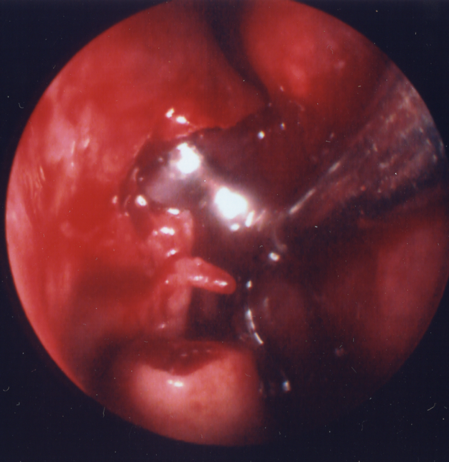
Figure 3. 9-mm endonasal balloon positioned in DCR ostium.
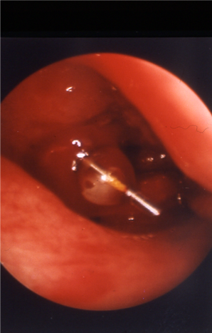
Figure 4. 5-mm endocanalicular balloon positioned at DCR ostium.
- Powered endoDCR
- Guarded electric burrs for osteotomy: 93% success in 283 cases (Ali, OPRS 2015)
- Ultrasonic endoscopic DCR with lacrimal stent: 93.1% anatomical and 88.6% functional success rate
- Can be useful in removing the thicker bone anteriorly
- Stentless endoscopic DCR
- Routine use of stents is very common, but there is evidence they might not be necessary. A number of conflicting studies have been published.
- In a randomized series of 128 eyes, success rates were higher in the stentless group of 65 eyes (89%) versus the stent group of 63 (57%) at 33 months (Mohamed, Ann R Coll Surg Engl 2013).
- Another study demonstrated no difference among 120 randomized patients (Chong, Ophthalmology 2013) and a third demonstrated 91% success among 132 cases without stents at 12 months and no new consecutive canalicular obstruction (Cannon, Ophthalmology 2013).
- A prospective, randomized, controlled interventional trial of 300 patients showed a statistically higher success rate in endonasal DCR surgery with tubes compared with no tubes. Failure rate was more than as twice as high when tubes were not used (12.2% vs. 5.3%) (Fayers, Ophthalmology 2016).
Ongoing controversies
- As endonasal/endoscopic surgical techniques have improved, success rates for endoDCR have also improved, and recent papers report 86%–99% success, compared to 85%–95% for external DCR.
- For ophthalmologists who are less familiar with nasal anatomy and instrumentation, initial endoDCRs might have lower success rates (Onerci, Acta Otolaryngol 2000).
- For experienced surgeons, OR time and blood loss are similar between the external and endoDCR techniques.
- Some patients and surgeons might prefer the conscious sedation anesthesia that can be used for external DCR but not usually for endonasal DCR (although Tripathi and Magab each report local anesthesia for endonasal DCR; see references).
- However, patients typically prefer the endonasal approach since there is no external scar, no dressing, no bruising, no sutures, and a more rapid return to work (Ozer, Int J Ophthalmol 2014; Gauba, Saudi J Ophthalmol 2014).
- One published study comparing external to nonlaser endoDCR by a single surgeon reported success rates of 90% and 89%, respectively, and noted that OR time was less for endoDCR (Dolman, Ophthalmology 2003).
- Patients with secondary causes of NLDO may have decreased success rates with endoDCR, in particular those with a history of radioactive iodine therapy and radiotherapy (Sweeney, Ophthalmic Plast Reconstr Surg 2018).
- Surgeons should discuss decreased success rates of endoDCR (as well as external DCR) with patients with a history of secondary causes of NLDO.
- Some value in the lack of disturbance to medial canthal anatomy and consequent zero risk of eyelid neurovascular injury from the endonasal approach.
- In cases of acute dacryocystitis for which an external incision might need to be delayed, the endonasal approach can be easily used during the acute infection and serves an important role for drainage of the abscess, shortening the overall course of the disease (Madge, Orbit 2011).
- Nevertheless, among ASOPRS members, external DCR is still preferred (Barmettler, Orbit 2013).
- Other ideas that may be considered controversial:
- Hyaluronic Acid (HA) nasal dressing application to the ostium: HA gel has been studied in the otolaryngology literature as an adjunct in endoscopic sinonasal surgery, its effectiveness remains controversial.
- In the functional endoscopic sinus surgery (FESS) literature, several studies found no significant reduction in synechiae formation when HA gel was compared with nonabsorbable gauze packing (Miller, Otolaryngol Head Neck Surg 2003) or with no postoperative dressing (Shi, Am J Rhinol Allergy 2013 and Wormald, Am J Rhinol 2006)
- In a single center, single surgeon, retrospective review of 254 patients who underwent endoDCR, intraop application of sodium hyaluronate solution (Protad) was associated with significantly higher anatomical (96.2% vs. 86.6%) and functional (93.3% vs. 85.2%) success rates compared with surgery without dressing. There was not a statistically significant difference in synechia formation, crusting and infection. (Park, Can J Ophthalmol 2017)
- In a randomized study of 161 patients with primary chronic dacryocystitis undergoing endoDCR, application of self-cross-linked HA hydrogel to the osteotomy site resulted in faster mucosal epithelialization, reduced granulation tissue formation, and improved long-term ostial patency compared with standard surgery alone. At 12 months, the HA hydrogel group achieved a significantly higher surgical success rate (89.0% vs. 77.2%, P < 0.05). (Yu, J Craniofac Surg 2021)
- Preservation of a nasal muscoal flap
- Some surgeons prefer to preserve nasal mucosa by elevating flap prior to creating osteotomy
- Semicircular incision made with a blade or periosteal elevator and folded back. Surgery performed as previously described.
- However, Zloto et al. conducted a retrospective review of 117 endoDCRs to compare outcomes with and without nasal mucosal flaps. They found no significant difference in surgical success between the two techniques (82.1% without flap vs. 86.8% with flap, P = 0.478), suggesting mucosal flap preservation did not improve endoDCR outcomes. (Zloto, Eye Lond. 2019)
Patient management: treatment and follow-up
- Do not blow nose for 2 weeks to reduce risk of postoperative epistaxis.
- Avoid manipulation of medial canthal area if a stent is placed to avoid prolapse laterally.
- Call surgeon or report to nearest ER for intractable epistaxis.
Medications prescribed:
- Consider topical ophthalmic antibiotic/steroid combination eyedrop for 1–2 weeks. Ointments may clog the outflow system.
- Consider nasal saline rinse followed by fluticasone or other nasal steroid for 1-3 months.
- Oral analgesia with acetaminophen, celecoxib or narcotic
Other management considerations:
- Patients commonly seen 2 weeks postoperatively for office nasal endoscopy and possible irrigation
- The Katena 23gauge bullet-tip cannula is easily fed into a canaliculus alongside the stent (if present); the typical relative ease of delivery into the nasopharynx (compared to preoperatively) is encouraging to the patient.
- Additional office follow-up at 6–8 weeks for nasal endoscopic exam: If the ostium is seen to be healing well without erythema and stenosis, the stent can be removed at 2–3 months or surgeon preference.
- Mucosal injections of triamcinolone can performed if aggressive fibrosis is seen.
- Some of the early enthusiasm for endoDCR techniques was related to reoperation for failing external DCR; many reports describe successful repeat endoDCR for ostium closure. However for reoperation, consider timing to avoid fibroblast proliferation, and possible use of mitomycin C or 5-FU.
Common treatment responses, follow-up strategies:
- Most patients will have a simple course with relief from epiphora and/or dacryocystitis. Sometimes, epiphora is further reduced upon removal of the silicone stent.
- Biofilm formation on the silicone stent has been described (Parsa, OPRS 2010) and although its clinical significance is not completely understood, it can sometimes cause blepharoconjunctivitis, increased ocular and/or nasal mucus discharge, and inflammation/stenosis of the ostium.
- Aggressive use of topical steroids and consideration of early removal of the stent (if ostium and/or canalicular inflammation do not abate) is indicated.
- Successful external DCR creates a 2–4mm ostium on average regardless of bony osteotomy size (Bumsted, Arch Otolaryngol 1982; Yazici, Arch Ophthalmol 2003). EndoDCR is likely to have similar results.
- Failing or failed endonasal DCR can occur from mucosal stenosis at the ostium. Repeat endonasal DCR (with or without antimetabolite) can be considered. Balloon dacryoplasty of the ostium for post-surgical stenosis has been reported to have 74% success (Lee, OPRS 2014).
Preventing and managing treatment complications
- Recurrent/progressive dacryostenosis:
- The literature is mixed on the question of ostium size: Size does not predict success (Chan, Ophthalmology 2013) versus a small ostium is associated with failure (Hammoudi, OPRS 2011).
- Associated with failure (Onerci, Acta Otolaryngol 2000):
- Bony spicules left at ostium
- Intranasal synechiae
- Inadequate removal of medial wall of the lacrimal sac
- Consider avoiding heat at the ostium, e.g., laser, Colorado needle (Garcia Vilaro, Ophthalmic Res 2013).
- Ostium granulomas recognized on postoperative endoscopy:
- Treat with intranasal steroid spray, triamcinolone injection, or excision (Ali, Orbit 2015).
- Sump syndrome with chronic mattering and low-grade dacryocystitis:
- Prevent with large uncinectomy and removal of medial wall of lacrimal sac (Fayet, OPRS 2014)
- Additionally it makes sense (although unproven) to prevent sump syndrome by extending the ostium to the inferior end of the lacrimal sac and preventing the flaps from reuniting by excising, suturing, etc.
- Cheese-wiring through puncta/canaliculi:
- Appropriate tension on silicone tubing loop in medial commissure necessary for prevention
- Avoid rigid silicone tubing such as Stent-Tube.
- Consider avoiding silicone intubation altogether (see stentless endoscopic DCR above).
- Perioperative dacryocystitis:
- Consider perioperative antibiotic treatment; not necessarily standard of care.
- Contact dermatitis from topical antibiotic drops:
- Neomycin and others can cause a reaction; stop if occurs.
- Intraoperative/postoperative hemorrhage
- Usual precautions regarding aspirin and anticoagulants.
- Multiple packing/nasal tampon options including resorbable nasal materials, Floseal, and nonabsorbable packing.
- Acute Sinusitis
- Typically occurs in the first postoperative week with usual symptoms of facial pain, tenderness and nasal discharge. Responds to oral antibiotics.
- Overall incidence in a retrospective cohort of 203 endoDCR cases was about 1.5%, except for patients with a history of chronic sinusitis, for whom the incidence was 15% (Shams, Eye 2013).
- Intranasal adhesions and synechiae:
- In-office lysis in first postoperative weeks
- Consider therapeutic (or prophylactic) use of resorbable or removable nasal tampon or packing to separate nasal septum and middle turbinate from the lateral nasal wall and ostium
- Cerebrospinal fluid (CSF) rhinorrhea:
- Uncommon; one case with spontaneous resolution on bedrest (Badilla, Arch Ophthalmol 2007)
- Prevent with careful bone removal, avoiding iatrogenic fracture, or removal of bone above the height of the medial canthal tendon.
- Consider early referral to ENT when suspected.
Historical perspective
- External DCR first described by Toti in 1904; previous lacrimal surgery for dacryocystitis described by Galen and Celsus involved using a “red-hot cautery”
- First balloon dacryoplasty described by Hanafee and Dayton in 1978
- Endonasal/endoscopic DCR
- First reported transnasal DCR by Caldwell in 1893; first modern clinical study published in J Laryngol Otol by McDonough and Meiring in 1989.
- In the ophthalmic literature, early reports with Holmium and other lasers were initially published by Massaro, Gonnering, and Harris in 1990.
References and additional resources
- American Society of Ophthalmic Plastic & Reconstructive Surgery, The Tear System. Accessed October 15, 2015.
- AAO, Basic and Clinical Science Course. Section 7: Orbit, Eyelids, and Lacrimal System, 2013-2014.
- AAO, Ophthalmic Technology Assessment: Endonasal Dacryocystorhinostomy, Ophthalmology. 2001:108:2369-2377.
- AAO, Preferred Practice Pattern, Nasolacrimal Duct Obstruction, 2013.
- Ali MJ et al. The Dacryocystorhinostomy Ostium Granulomas: Classification, Indications for Treatment, Management Modalities and Outcomes. Orbit. 2015 Jun;34(3):146-51
- Ali MJ, Psaltis AJ, Murphy J, Wormald PJ. Powered endoscopic dacryocystorhinostomy: a decade of experience. Ophthal Plast Reconstr Surg. 2015; 31(3):219-221.
- Ali MJ, Singh M, Chisty N, Kamal S, Naik MN. Endoscopic ultrasonic dacryocystorhinostomy: clinical profile and outcomes. Eur Arch Otorhinolaryngol. 2016 Jul;273(7):1789-93. doi: 10.1007/s00405-015-3826-z. Epub 2015 Nov 3. PMID: 26530294.
- Badilla J, Dolman PJ. Cerebrospinal fluid leaks complicating orbital or oculoplastic surgery. Arch Ophthalmol. 2007; 125(12):1631-1634.
- Barmettler A, Ehrlich JR, Lelli G Jr. Current preferences and reported success rates in dacryocystorhinostomy amongst ASOPRS members. Orbit. 2013; 32(1):20-6.
- Bumsted RM, Linberg JV, Anderson RL, Barreras R. External dacryocystorhinostomy. A prospective study comparing the size of the operative and healed ostium. Arch Otolaryngol. 1982; 108(7):407-410.
- Camara JG, Bengzon AU, Henson RD. The safety and efficacy of mitomycin C in endonasal endoscopic laser-assisted dacryocystorhinostomy. Ophthal Plast Reconstr Surg. 2000;16(2):114-118.
- Cannon PS, Chan W, Selva D. Incidence of canalicular closure with endonasal dacryocystorhinostomy without intubation in primary nasolacrimal duct obstruction. Ophthalmology. 2013; 120(8):1688-1692.
- Chan W, Selva D. Ostium shrinkage after endoscopic dacryocystorhinostomy. Ophthalmology. 2013; 120(8):1693-1696.
- Chong KK, Lai FH, Ho M, Luk A, Wong BW, Young A. Randomized trial on silicone intubation in endoscopic mechanical dacryocystorhinostomy (SEND) for primary nasolacrimal duct obstruction. Ophthalmology. 2013; 120(10):2139-2145.
- Dolman PJ. Comparison of external dacryocystorhinostomy with nonlaser endonasal dacryocystorhinostomy. Ophthalmology. 2003;110(1):78-84.
- Dolmetsch AM. Nonlaser endoscopic endonasal dacryocystorhinostomy with adjunctive mitomycin C in nasolacrimal duct obstruction in adults. Ophthalmology. 2010; 117(5):1037-1040.
- Fayers T, Dolman PJ. Bicanalicular silicone stents in endonasal dacryocystorhinostomy: results of a randomized clinical trial. Ophthalmology. 2016;123(10): 2255-9.
- Fayet B, Katowitz WR, Racy E, Ruban JM, Katowitz JA. Endoscopic dacryocystorhinostomy: the keys to surgical success. Ophthal Plast Reconstr Surg. 2014; 30(1):69-71.
- Fenton S, Cleary PE, Horan E, Murray A, Ho SL, Ryder D, O’Connor G. Balloon dacryocystoplasty study in the management of adult epiphora. Eye (Lond). 2001; 15(Pt 1):67-69.
- Garcia Vilaro M et al. Thermal damage influences endonasal dacryocystorhinostomy success. Ophthalmic Res. 2013; 49(4):209-214.
- Gauba V. External versus endonasal dacryocytorhinostomy in a specialized lacrimal surgery center. Saudi J Ophthalmol. 2014; 28(1):36-9.
- Gustafson RO, Bartley GB. A plea for preservation of the middle turbinate during dacryocystorhinostomy. Ophthal Plast Reconstr Surg. 1999; 15(2):75-76.
- Hammoudi DS, Tucker NA. Factors associated with outcome of endonasal dacryocystorhinostomy. Ophthal Plast Reconstr Surg. 2011; 27(4):266-269.
- Hodgson N, Bratton E, Whipple K, Priel A, Oh SR, Fante RG, Kikkawa DO, Korn BS. Outcomes of endonasal dacryocystorhinostomy without mucosal flap preservation. Ophthal Plast Reconstr Surg. 2014; 30(1):24-27.
- Jang SY, Lee KH, Lee SY, Yoon JS. Effects of nasopore packing on dacryocystorhinostomy. Korean J Ophthalmol. 2013; 27(2):73-80.
- Jutley G et al. Patient satisfaction following endoscopic endonasal dacryocystorhinostomy: a quality of life study. Eye (Lond). 2013; 27(9):1084-1089.
- Kamal S, Ali MJ, Naik MN. Circumostial injection of mitomycin C (COS-MMC) in external and endoscopic dacryocystorhinostomy: efficacy, safety profile, and outcomes. Ophthal Plast Reconstr Surg. 2014; 30(2):187-190.
- Kao SC, Liao CL, Tseng JH, Chen MS, Hou PK. Dacryocystorhinostomy with intraoperative mitomycin C. Ophthalmology. 1997; 104(1):86-91.
- Kuchar A, Steinkogler FJ. Antegrade balloon dilatation of nasolacrimal duct obstruction in adults. Br J Ophthalmol. 2001;85(2):200-204.
- Lanciego C, Toledano N, De Miguel S, Perea M, Padilla M, Rodriguez-Merlo R, Dávila J, Ibarburen C, Cano C, García IR, García LG. Resolution of epiphora with nasolacrimal stents: results of long-term follow-up in a multicenter prospective study. J Vasc Interv Radiol. 2003; 14(11):1417-1425.
- Lee A, Ali MJ, Li EY, Wong AC, Yuen HK. Balloon dacryoplasty in internal ostium stenosis after endoscopic dacryocystorhinostomy. Ophthal Plast Reconstr Surg. 2014; 30(1):7-10.
- Madge SN, Chan W, Malhotra R, Ghabrial R, Floreani S, Wormald PJ, Tsirbas A, Selva D. Endoscopic dacryocystorhinostomy in acute dacryocystitis: a multicenter case series. Orbit. 2011; 30(1):1-6.
- Maini S, Raghava N, Youngs R, Evans K, Trivedi S, Foy C, Mackintosh G. Endoscopic endonasal laser versus endonasal surgical dacryocystorhinostomy for epiphora due to nasolacrimal duct obstruction: prospective, randomised, controlled trial. J Laryngol Otol. 2007; 121(12):1170-1176.
- Marcet MM, Kuk AK, Phelps PO. Evidence-based review of surgical practices in endoscopic endonasal dacryocystorhinostomy for primary acquired nasolacrimal duct obstruction and other new indications. Curr Opin Ophthalmol. 2014; 25(5):443-8.
- McCullough KM. Naso-lacrimal duct balloon dilatation: medium to long term follow-up. Clin Radiol. 2001; 56(1):13-16.
- Miller RS, Steward DL, Tami TA, et al. The clinical effects of hyaluronic acid ester nasal dressing (Merogel) on intranasal wound healing after functional endoscopic sinus surgery. Otolaryngol Head Neck Surg. 2003;128(6):862-869.
- Mohamed SH, Khan I, Shakeel M, Nandapalan V. Long-term results of endonasal dacryocystorhinostomy with and without stenting. Ann R Coll Surg Engl. 2013; 95(3):196-199.
- Moore WM, Bentley CR, Olver JM. Functional and anatomic results after two types of endoscopic endonasal dacryocystorhinostomy: surgical and holmium laser. Ophthalmology. 2002; 109(8):1575-1582.
- Nagi KS, Meyer DR. Utilization patterns for diagnostic imaging in the evaluation of epiphora due to lacrimal obstruction: a national survey. Ophthal Plast Reconstr Surg. 2010; 26(3):168-171.
- O’Donnell B, Shah R. Dacryocystorhinostomy for epiphora in the presence of a patent lacrimal system. Clin Experiment Ophthalmol. 2001; 29(1):27-29.
- O’Donnell B, Clement CI. Assessing patients with epiphora who are patent to syringing: clinical predictors of response to dacryocystorhinostomy. Ophthal Plast Reconstr Surg. 2007; 23(3):173-178.
- Onerci M, Orhan M, Ogretmenoğlu O, Irkeç M. Long-term results and reasons for failure of intranasal endoscopic dacryocystorhinostomy. Acta Otolaryngol. 2000; 120(2):319-322.
- Ozer S, Ozer PA. Endoscopic vs external dacryocystorhinostomy-comparison from the patients’ aspect. Int J Ophthalmol. 2014; 7(4):689-696.
- Park J, Lee J, Jang S, et al. Effectiveness of sodium hyaluronate (Protad) application in endoscopic endonasal dacryocystorhinostomy. Can J Ophthalmol. 2017;52(2):192-197.
- Parsa K, Schaudinn C, Gorur A, Sedghizadeh PP, Johnson T, Tse DT, Costerton JW. Demonstration of bacterial biofilms in culture-negative silicone stent and jones tube. Ophthal Plast Reconstr Surg. 2010; 26(6):426-430.
- Patel V, Ross JJ, Malhotra R. Early experience using a new modified bone nibbler for superior osteotomy during endonasal dacryocystorhinostomy. Ophthal Plast Reconstr Surg. 2011; 27(1):15-20.
- Phelps PO, Abariga SA, Cowling BJ, Selva D, Marcet MM. Antimetabolites as an adjunct to dacryocystorhinostomy for nasolacrimal duct obstruction. Cochrane Database Syst Rev. 2020 Apr 7;4(4):CD012309.
- Ragab SM, el-Koddousy MS, Badr M. Endocanalicular, high-pressure balloon catheter, endoscopic dacryocystorhinostomy: a randomized controlled trial. Otolaryngol Head Neck Surg. 2011; 145(4):683-688.
- Shams PN et al. Acute post-operative rhinosinusitis following endonasal dacryocystorhinostomy. Eye (Lond). 2013; 27(10):1130-1136.
- Shams PN, Wormald PJ, Selva D. Anatomical landmarks of the lateral nasal wall: implications for endonasal lacrimal surgery. Curr Opin Ophthalmol. 2015; 26(5):408-15.
- Shi R, Zhou J, Wang B, et al. The clinical outcomes of new hyaluronan nasal dressing: a prospective, randomized, controlled study. Am J Rhinol Allergy. 2013;27(1):71-76.
- Silbert DI, Matta NS. Outcomes of 9 mm balloon-assisted endoscopic dacryocystorhinostomy: retrospective review of 97 cases. Orbit. 2010; 29(3):131-134.
- Sinha AK, Kahana EL, Meijome TE, Jacobson A, Spadaro JZ, Chung SY, Kahana A. Successful outcomes of pediatric dacryocystorhinostomy with adjunctive 5-fluorouracil. Front Ophthalmol (Lausanne). 2026 Jul 17;6:1805226.
- Sweeney AR, Davis GE, Chang SH, Amadi AJ. Outcomes of endoscopic dacryocystorhinostomy in secondary acquired nasolacrimal duct obstruction: a case-control study. Ophthalmic Plast Reconstr Surg. 2018; 34(1):20-25.
- Tripathi A, Lesser TH, O’Donnell NP, White S. Local anaesthetic endonasal endoscopic laser dacryocystorhinostomy: analysis of patients’ acceptability and various factors affecting the success of this procedure. Eye (Lond). 2002; 16(2):146-149.
- Trotter WL, Meyer DR. Endoscopic conjunctivodacryocystorhinostomy with Jones tube placement. Ophthalmology. 2000; 107(6):1206-1209.
- Tsai CC et al. Efficacy of probing the nasolacrimal duct with adjunctive Mitomycin-C for epiphora in adults. Ophthalmology. 2002; 109(1):172-174.
- Ugurbas SH, Zilelioglu G, Sargon MF, Anadolu Y, Akiner M, Aktürk T. Histopathologic effects of mitomycin-C on endoscopic transnasal dacryocystorhinostomy. Ophthalmic Surg Lasers. 1997; 28(4):300-304.
- Velegrakis GA, Prokopakis EP, Panayotaki I, Pagalos AG, Siganos CS, Helidonis ES. Intranasal laser-assisted dacryocystorhinostomy with the use of a surgical microscope. Am J Otolaryngol. 2002; 23(5):272-276.
- Watkins LM, Janfaza P, Rubin PA. The evolution of endonasal dacryocystorhinostomy. Surv Ophthalmol. 2003;48:73-84. Comment in: Surv Ophthalmol. 2004;49:543; author reply 544.
- Weiss M, Lauer SA, Fried MP, Uribe J, Sadoughi B. Endoscopic endonasal surgery simulator as a training tool for ophthalmology residents. Ophthal Plast Reconstr Surg. 2008; 24(6):460-464.
- Wormald PJ, Boustred RN, Le T, Hawke L, Sacks R. A prospective single-blind randomized controlled study of use of hyaluronic acid nasal packs in patients after endoscopic sinus surgery. Am J Rhinol. 2006;20(1):7-10.
- Xue K, Mellington FE, Norris JH. Meta-analysis of the adjunctive use of mitomycin C in primary and revision, external and endonasal dacryocystorhinostomy. Orbit. 2014; 33(4):239-244.
- Yazici B, Yazici Z. Final nasolacrimal ostium after external dacryocystorhinostomy. Arch Ophthalmol. 2003; 121(1):76-80.
- Yu B, Tu Y, Zhou G, Shi J, Wu E, Wu W. Self-Cross-Linked Hyaluronic Acid Hydrogel in Endonasal Endoscopic Dacryocystorhinostomy: A Randomized, Controlled Trial. Journal of Craniofacial Surgery. 2021; 32 (5): 1942-1945.
- Ziahosseini K, Al-Abbadi Z, Malhotra R. Botulinum toxin injection for the treatment of epiphora in lacrimal outflow obstruction. Eye. 2015; 29(5):656-61.
- Zloto O, Koval T, Yakirevich A, et al. Endoscopic dacryocystorhinostomy with and without mucosal flap-is there any difference?. Eye (Lond). 2020;34(8):1449-1453.
Financial disclosures
Reviewers
Dianne Schlachter: None