Anatomy of the External Ear
Updated May 2024
Anatomic units

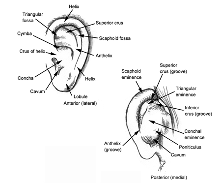
Figures 1 and 2. Anatomy of the external ear. External ear superficial landmarks, medially and laterally.
- The external ear has a cartilaginous framework in all areas except the lobule.
- The outer curvature of the superior half of the auricle is the helix and is concave laterally.
- The antihelix is the next structure inward and is concave medially.
- The antihelix divides into an inferoanterior crus and a superoposterior crus that parallels the helix.
- The space between the helix and the antihelix is known as the scaphoid fossa.
- The space between the crus of the antihelix is known as the triangular fossa.
- The space between the inferoanterior crus and the anterior terminus of the helix is known as the cymba.
- Failure of the antihelix to form properly is one cause of prominent ears and the “lop ear” deformity.
- Inferiorly, the two crus unite to form the posterior boundary of the conchal bowl.
- The concha is the hollow depression in the central part of the auricle that directs sound into the external auditory meatus, with which it is continuous.
- The concha is concave laterally.
- An excessively deep conchal bowl projects the ear away from the mastoid surface and results in a more prominent ear.
- The external auditory canal is a sigmoid shaped tube lined with skin and cartilage and supported by the temporal bone. It terminates at the tympanic membrane. Its initial direction is superoanterior and it ends in a inferoanterior direction.
- The tragus is the flap of tissue at the anterior border of the concha that can be digitally depressed to close the external auditory meatus.
- Opposite the tragus is the antitragus forming the inferior rim of the concha.
- The lobule hangs from the antitragus and is variably attached anteriorly to the skin superior to the mandibular angle.
- The medial (posterior) landmarks are less familiar and include the conchal eminence and the grooves created by the two crus of the antihelix
- The angles of attachment of the ear to the head can be measured and used to define normal (Aesthetic Plast Surg 2008).
- Scaphaconchal (normal avg 107 degrees)
- Cephaloauricular (normal avg 31 degrees)
- Auricular cartilage forms an incomplete skeleton for the visible auricle that is augmented with connective tissue and muscle.
- Cutaneous innervation of the external ear (Figure 3)

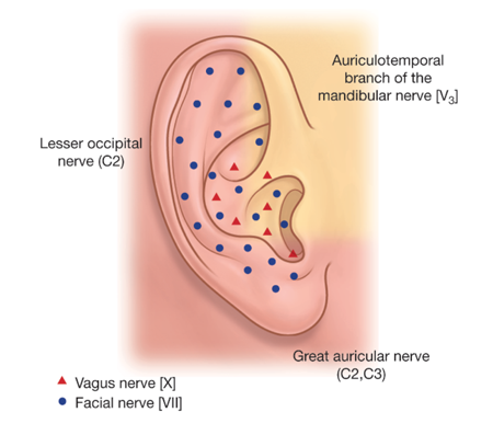
Figure 3. Sensory innervation of the external ear.
- Greater auricular nerve from the cervical plexus (C2, C3) innervates the lobule and the inferior portions of the helix, antihelix, and concha.
- Lesser occipital nerve from the cervical plexus (C2) innervates the superoposterior portion of the auricle.
- Auriculotemporal branch of the mandibular branch (V3) of the trigeminal supplies the tragus, anterior helix, and the external auditory meatus.
- Auricular branch of the vagus nerve (CN X) innervates the deeper parts of the auricle adjacent to the external auditory canal.
- Involuntary cough can result from cleaning of the external ear for certain individuals.
- The facial nerve (CN VII) can also contribute to the auricular branch of the vagus.
- Vascular supply of the external ear is from branches of the external carotid.
- Posterior auricular artery (a.) and vein (v.)
- Superficial temporal a. and v.
- Occipital a. and v.
- Lymphatic drainage
- Anteriorly to the parotid nodes
- Posteriorly to the mastoid nodes and upper deep cervical nodes
- Intrinsic and extrinsic muscles of the external ear
- Intrinsic muscles pass among the cartilaginous portions of the auricle and can change its shape.
- Extrinsic muscles arise from the scalp or skull anteriorly, posteriorly, and superiorly to the auricle and insert variably on the cartilaginous structure to change ear position.
- The auricularis posterior is the most developed of these and inserts on the medial surface of the concha. This muscle arises from the mastoid fascia and lies anterior and lateral to the insertion of the sternocleidomastoid muscle.
- Relevant clinical correlations
- Grafts of pre- and postauricular skin have been described for eyelid reconstruction.
- The lateral surface of the auricle is visible to onlookers, and so the medial surface (the “back of the ear”) is most commonly used for external ear surgical incisions and harvest of cartilage or skin grafts.
- The most commonly type of otoplasty is directed at reforming an underdeveloped antitragus to repair the “lop ear” deformity and reduce the prominence of the auricles.
Historical perspective
The Edwin Smith Surgical Papyrus (c. 1600 BCE) references vessels to the ears: two to the right ear are said to carry the “breath of life” and two to the left the “breath of death.”
References and additional resources
- Da Silva Freitas R et al. Comparing cephaloauricular and scaphaconchal angles in prominent ear patients and control subjects. Aesthetic Plast Surg 2008; 32(4):620-3.
- Drake RL, Vogl AW, Mitchell AWM. Gray’s Anatomy for Students, 2nd Edition. Elsevier 2009. (Accessed at https://www.inkling.com/read/gray-anatomy-students-drake-vogl-mitchell-2nd/chapter-8/regional-anatomy-ear)
- Lin SJ. Prominent Ear. Medscape 2014. (Accessed at http://emedicine.medscape.com/article/1290275-overview#showall)
