Tinea faciei
Updated May 2025
Alexis Kassotis and Lora R. Dagi Glass, MD
Tinea infections are defined by their anatomic location. This review will focus on the periocular manifestations of tinea faciei. Tinea faciei, by definition, involves only the glabrous skin of the face. Periocular tinea infection involving the eyebrow is a manifestation of tinea capitis while tinea of the bearded area of the face is known as tinea barbae.
Establishing the diagnosis
Etiology
- Fungal infection of the superficial skin.
- Caused by dermatophytes which require keratin for growth.
- Trichophyton tonsurans and T. rubrum, (harbored by humans) as well as Microsporum canis (harbored by animals) are the most common infectious species.
- Worldwide, rates of tinea faciei due to T. interdigitale are increasing.
- Acquired by direct contact with infected humans or, less commonly, animals.
- Can also be acquired indirectly by exposure to fomites or fungi-laden soil.
Epidemiology
- Tinea faciei accounts for approximately 4% of tinea corporis, a general term for tinea infection manifesting anywhere on the glabrous skin of the face (Lin, 2004).
- Tinea corporis is sometimes misleadingly called “ringworm,” because of its distinctive, slightly raised, ring-shaped rash.
- Tinea blepharociliaris is the more specific term when infection involves the eyelashes and eyelids (Nguyen, 2023; Martínez-Ortega, 2024).
- Disease is most likely to present from early childhood to the fourth decade of life (Lin, 2004).
- Young children are likely to acquire tinea faciei from contact with domesticated animals or other children.
- Tinea faciei has been reported in neonates as early as day 2 of life (Alkeswani, 2018).
- Tinea infections account for nearly 30% of skin infections in high school athletes due to their contact with fungal-laden athletic equipment, locker rooms and showers. One-fourth of these cases occur on the head or face (Ashack, 2016).
- Occlusive clothing, living in a hot humid environment, poor hygiene, proximity to animals, and crowded living conditions are important risk factors (Hill, 2024).
- Tinea faciei is the most commonly misdiagnosed cutaneous fungal infection and it is particularly underdiagnosed in the periocular region (Basak, 2011 & Lin, 2004).
- A retrospective study found that the majority of cases were not diagnosed until 1-4 months after symptom onset (Aste, 2009).
- Misdiagnosis is due to its highly polymorphic and sometimes subtle presentation.
- This is problematic as misdiagnosis leads to inappropriate treatment with corticosteroids, which can worsen disease, alter presentation and hinder diagnosis (see clinical presentation and management).
- Tinea incognita is the term given to a fungal disease altered in appearance by incorrect treatment (i.e. topical corticosteroids).
- This is problematic as misdiagnosis leads to inappropriate treatment with corticosteroids, which can worsen disease, alter presentation and hinder diagnosis (see clinical presentation and management).
- For the above reasons, the true incidence of tinea faciei is difficult to determine.
Clinical features
- Classical presentation includes single or multiple annular lesions with central clearing and scale formation (Figure 1).
- May be acute in onset with rapid extension or subacute/chronic with slow spread.
- Often an erythematous, inflamed rim around the rash, known as the active border.
- A periocular rash accompanied by eyelash and/or eyebrow hair loss is highly specific for tinea faciei.
- Other presenting features include:
- Periocular tenderness
- Periocular edema
- Pruritus and burning which is worse after sun exposure
- Lymphadenopathy
- Lesion alteration with the use of topical steroids
- Steroids reduce erythema and inflammation and may lead to changes in skin pigmentation, skin thinning and striae formation. This modified clinical appearance makes diagnosis more difficult.
- Atypical features are observed in approximately 40% of cases and include (Aste, 2009):
- Confluent erythematous plaques
- Vesicles and/or bullae
- Papules and/or pustules
- Tinea faciei on the eyelid mimicking hordeolum has been reported (Yin, 2018).
Other diagnostic tests (often required for diagnosis)
- Microscopic examination of skin scrapings in 10% potassium hydroxide (KOH) solution
- Woods lamp examination
- Fungal culture
- When feasible, mycological confirmation before starting treatment is considered best practice (Hill, 2024)
- Can provide specific strain and antifungal sensitivities
- May take several days to weeks for growth
- When feasible, mycological confirmation before starting treatment is considered best practice (Hill, 2024)
- False negative rate of fungal culture is up to 30% (Lin, 2004).
- Histologic examination of tissue
- Periodic acid–Schiff (PAS) staining is key to ruling out other dermatoses.
- Hyphae in the stratum corneum is specific to tinea infection and seen with PAS staining.
- Periodic acid–Schiff (PAS) staining is key to ruling out other dermatoses.
- Dermatology consult should be considered for atypical presentations.
Differential diagnosis
- Eczema
- Chronic blepharitis
- Impetigo
- Acne rosacea
- Seborrheic dermatitis
- Periorificial dermatitis
- Contact dermatitis
- Herpes simplex virus
- Actinic keratoses
- Cutaneous lupus erythematosus
- Granuloma annulare
- Cutaneous leprosy
- Erythema annulare centrifugum
- Erythema migrans
Patient management: treatment and follow-up
- Medical therapy:
- Topical antifungal cream is first line for tinea faciei.
- Commonly used regimens are topical azoles (i.e. ketoconazole).
- Continued for 2 weeks beyond clinical resolution (Rengasamy, 2020).
- Oral medications such as griseofulvin, itraconazole or terbinafine may be used.
- if disease is non-responsive to topical treatment .
- If disease infiltrates past the superficial cutaneous layers (i.e. into the vellus hair follicles of the face).
- If disease involves eyelid margin/eyelashes.
- Trichophyton indotneae species is resistant to terbinafine, sensitive to itraconazole.
- Prevalent strain of Indian subcontinent (Uhrlaß, 2022).
- Long duration of treatment (weeks to months) is often required to ensure complete eradication of fungi.
- In some cases, oral and topical antifungals are used in combination.
- Corticosteroids are contraindicated as they do not eradicate fungi from the skin surface and are associated with:
- Recurrence
- Spread of disease
- Antifungal resistance
- Surgical therapy: none
- Prognosis: complete resolution with hair restoration takes approximately 3 months with appropriate treatment.
- Dermatology consult should be considered for difficult to treat or refractory cases.
- Good hygiene practices are important:
- Regular washing and thorough drying of skin important to prevent the spread of infection.
- Avoid sharing towels, clothing and other personal items with others to prevent spread or recurrence.
Complications
- Inadequate eradication of infection leading to chronic recurrence.
- In rare cases, the initial infection site can be asymptomatic, leading to chronic infection at another site(s).
- A child with a decade of recurrent tinea faciei involving the eyelid was found to be an asymptomatic carrier of tinea capitis, with repeated autoinoculation of the face (Kawachi, 2010)
- Scarring
- Kerion (formation of a fungal abscess)
- Adverse effects of treatment:
- Griseofulvin
- Nausea
- Vomiting
- Photosensitivity
- Terbinafine
- Liver toxicity
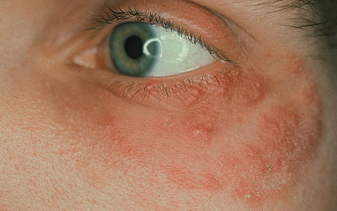
Photograph courtesy of D@nderm Atlas of Clinical Dermatology.
Figure 1. Periocular tinea faciei due to infection with Trichophyton mentagrophytes. An erythematous annular rash with central clearing and edema of the upper eyelid can be seen.
References and additional resources
- Andrews M. Burns M. Common tinea infections in children. Am Fam Physician. 2008 May 15;77(10):1415-1420.
- Cirillo-Hyland V, Humphreys T, Elenitsas R. Tinea faciei. J Am Acad Dermatol. 1993;29(1):119-120.
- Hill RC, Caplan AS, Elewski B, Gold JAW, Lockhart SR, Smith DJ, Lipner SR. Expert Panel Review of Skin and Hair Dermatophytoses in an Era of Antifungal Resistance. Am J Clin Dermatol. 2024 May;25(3):359-389.
- Lin RL, Szepietowski JC, Schwartz RA. Tinea faciei, an often-deceptive facial eruption. International Journal of Dermatology. 2004; 43(6):437-449.
- Nguyen B, Hu JK, Tosti A. Eyebrow and Eyelash Alopecia: A Clinical Review. Am J Clin Dermatol. 2023 Jan;24(1):55-67.
- Martínez-Ortega JI, Mut Quej JE, Medina Angulo TK. Tinea Blepharociliaris: A Case Report and Literature Review. Cureus. 2024 Dec 24;16(12):e76308.
- Alkeswani A, Duncan JR, Theos A. Tinea faciei starting at day two of life. Pediatr Dermatol. 2019;36(1):e20‐e22.
- Ashack KA, Burton KA, Johnson TR, Currie DW, Comstock RD, Dellavalle RP. Skin infections among US high school athletes: A national survey. J Am Acad Dermatol. 2016;74(4):679‐84.e1.
- Basak SA, Berk DR, Lueder GT, Bayliss SJ. Common Features of Periocular Tinea. Arch Ophthalmol. 2011;129(3):306–309.
- Yin B, Ran X, Ran Y, Zhang Y, Pradhan S. Cover Image: Dermoscopic detection of unusual eyelash Trichophyton interdigitale infection mimicking hordeolum. Br J Dermatol. 2018;178(4):989‐990.
- Aste N, Atzori L, Aste N, Pau M. A 20-year survery of tinea faciei. Mycoses. 2009;53(6):504-508.
- Rademaker M, Havill S. Griseofulvin and terbinafine in the treatment of tinea capitis in children. The New Zealand Medical Journal. 1998 Feb;111(1060):55-57.
- Rengasamy M, Shenoy MM, Dogra S, et al. Indian Association of Dermatologists, Venereologists and Leprologists (IADVL) Task Force against Recalcitrant Tinea (ITART) Consensus on the Management of Glabrous Tinea (INTACT). Indian Dermatol Online J. 2020 Jul 13;11(4):502-519.
- Uhrlaß S, Verma SB, Gräser Y, Rezaei-Matehkolaei A, Hatami M, Schaller M, Nenoff P. Trichophyton indotineae-An Emerging Pathogen Causing Recalcitrant Dermatophytoses in India and Worldwide-A Multidimensional Perspective. J Fungi (Basel). 2022 Jul 21;8(7):757.
- Kawachi Y, Ikegami M, Takase T, Otsuka F. Chronically Recurrent and Disseminated Tinea Faciei/Corporis—Autoinoculation from Asymptomatic Tinea Capitis Carriage. Ped Dermatol. 2010;27(5):527-528.
- Majid I, Sheikh G, Kanth F, Hakak R. Relapse after Oral Terbinafine Therapy in Dermatophytosis: A Clinical and Mycological Study. Indian J Dermatol. 2016 Sep-Oct;61(5):529-33.
- Khiewplueang K, Leeyaphan C, Bunyaratavej S, Jirawattanadon P, Saengthong-Aram P, Matthapan L, Prasong W, Panyawong C, Plengpanich A. Tinea faciei clinical characteristics, causative agents, treatments and outcomes; a retrospective study in Thailand. Mycoses. 2024 Jun;67(6):e13754.
- Verma S. Steroid Modified tinea. BMJ. 2017;356:j973
- Hainer B. Dermatophyte infections. Am Fam Physician. 2003 Jan 1;67(1):101-109.
- del Boz J, Crespo V, de Troya M. Pediatric Tinea Faciei in Southern Spain: A 30‐Year Survey. Ped Dermatol. 2011; 29(3): 249-253.
- Amigo M, Milani-Nejad N, Mosser-Goldfarb J. Periocular Tinea Faciei. J Pediatr. 2020 Jun;221:255-256.
Financial disclosures
Reviewers
Dianne Schlachter – Consultant & Speaker, Amgen