Generalized staphylococcal scalded skin syndrome
Alexis Kassotis and Lora R. Dagi Glass, MD
Establishing the diagnosis
Etiology
- Staphylococcal scalded skin syndrome (SSSS) is a potentially lethal dermatologic condition.
- It is caused by strains of Staphylococcus aureus that produce exfoliative exotoxins A and B (ETA and ETB).
- These exotoxins act as serine proteases that cleave desmoglein 1 (a desmosomal protein); cleavage leads to loss of keratinocyte-keratinocyte adhesion in the stratum granulosum, which compromises skin architecture.
- Rates of SSSS due to Methicillin-Resistant S. Aureus (MRSA) vary by location but are relatively low.
- It is caused by strains of Staphylococcus aureus that produce exfoliative exotoxins A and B (ETA and ETB).
- There are two forms of SSSS:
- Localized disease (known as bullous impetigo), which rarely affects the periocular region.
- Generalized disease, which almost always affects the periocular region.
- This review will focus on the generalized form of SSSS, which occurs when the exotoxin spreads hematogenously, causing widespread damage to the epidermis.
Epidemiology
- Estimated incidence is between 0.09 and 0.56 cases per million (Handler, 2014).
- Primarily a disease of immunocompetent children
- Disease most commonly occurs from the neonatal period (as early as 48 hours after birth) to 5 years of age.
- Antibodies against the staphylococcal exotoxins are typically acquired in childhood, which accounts for the relative rarity of disease in older children and adults.
- Infants are especially vulnerable due to immature renal function, leading to reduced clearance of exotoxins.
- Approximately one-third of healthy individuals are colonized with S. Aureus and can serve as a vector of disease (Sakr, 2018).
- Outbreaks are commonly seen in schools and day care facilities.
- Disease most commonly occurs from the neonatal period (as early as 48 hours after birth) to 5 years of age.
- SSSS is rare in adults; about 60 cases have been documented.
- Renal impairment is a major risk factor as it leads to impaired excretion of circulating exotoxins. However, multiple cases have been reported in immunocompetent adults without renal dysfunction (Patel, 2000).
Clinical features
- History
- Prodrome of fever, agitation and irritability (particularly in young children), sore throat, anorexia, and conjunctivitis.
- After a non-specific prodromal period, erythroderma develops. This is followed by superficial desquamation with some areas of skin sparing. This gives the skin a wrinkled appearance and skin may slough off with gentle pressure (Nikolsky sign). After this, fragile vesicles and bullae form that then burst to reveal an erythematous base.
- Periocular crusting often occurs after lesions burst.
- Disease is confined to the epidermis which differentiates SSSS from Toxic epidermal necrolysis (TEN), a common mimic characterized by dermal involvement.
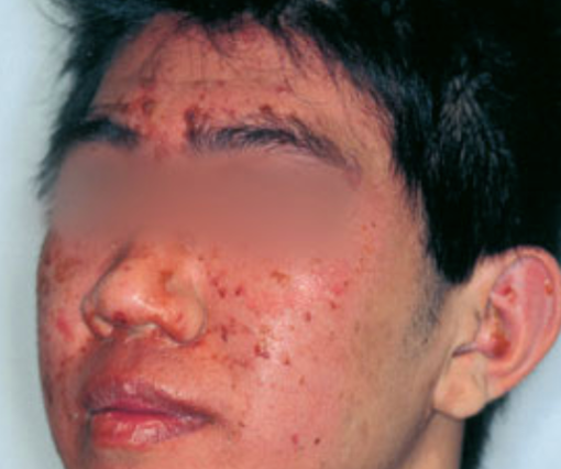
- Disease classically begins on the head or face (Figure 1).
- In neonates, lesions are typically also present around the umbilical stump and in the diaper area.
- Absence of mucous membrane involvement helps differentiate SSSS from similar blistering conditions including TEN and Stevens-Johnson syndrome.
- Systemic features including fever, malaise, and signs of significant dehydration such as poor skin turgor and delayed capillary refill time are usually present.
Other diagnostic tests (required)
- Typical clinical pattern (see clinical features)
- Skin biopsy and histopathologic evaluation demonstrating:
- Detachment of the superficial epidermis beginning at the stratum granulosum.
- Lack of notable inflammation.
- Isolation of S. Aureus producing ETA or ETB
- Skin cultures with nasal swab
- Blood cultures
- Culture of the umbilical stump in neonates
Other diagnostic studies
- Tzanck smear
- May be combined with negative direct immunofluorescence to increase specificity.
- Gram stain (demonstrating clusters of gram-positive cocci) may be used in a resource limited setting.
- In conjunction with typical clinical features, suggestive findings on gram stain is justification to start empiric therapy.
- Testing is relatively rapid; thus, it may shorten time between presentation and treatment.
- As disease is toxin mediated, samples cannot be taken from bullae or vesicles as they will not contain bacteria.
- Dermatoscopy demonstrates erosions with peripheral epidermal remnants and can help support the diagnosis.
Differential diagnosis
- Toxic epidermal necrolysis
- Stevens-Johnson Syndrome
- Epidermolysis bullosa
- Pemphigus foliaceous
- Drug reaction with eosinophilia and systemic symptoms (DRESS)
- Staphylococcal toxic shock syndrome
Patient management: treatment and follow-up
- Medical therapy: treatment is multidisciplinary and often requires a burn unit or intensive care unit.
- Early treatment with intravenous anti-staphylococcal antibiotics (i.e. beta-lactamase resistant penicillin) is critical in decreasing morbidity and mortality.
- Flucloxacillin is a commonly used for adults and children.
- Topical mupirocin may be used to eradicate nasal colonization of S. Aureus and topical antibiotics can be added for concurrent conjunctivitis.
- Clarithromycin or cefuroxime can be used in penicillin allergy.
- Adjuvant clindamycin, vancomycin or linezolid may be used if MRSA is suspected.
- MRSA coverage is generally added empirically in areas of high MRSA prevalence or if patients fail to improve with standard therapy.
- However, compared to Staphylococcal infections overall, those associated with SSSS are less likely to be methicillin-resistant and more likely to be clindamycin-resistant.
- The combination of clindamycin with melittin (a drug with broad spectrum antibacterial, antiviral, antifungal, antiprotozoal and anti-inflammatory effects) has been recently demonstrated to have synergistic activity against Staphylococcal exfoliative exotoxin A and B (Mahmoudi, 2020).
- Fresh frozen plasma (FFP) was demonstrated in a case series to effectively neutralize antibodies against exotoxin A in the pediatric population (Tenendbaum, 2007).
- Use of intravenous immunoglobulin (IVIG) can be attempted in children who have failed other treatments, although its use may be associated with prolonged hospitalization (Li, 2013 & Handler, 2014).
- A recent case report demonstrated the efficacy of IVIG as part of a multimodal treatment regimen in an adult with SSSS (Urata, 2018).
- Analgesics (i.e. acetaminophen, fentanyl)
- Intravenous fluid resuscitation
- Nasogastric feeding
- Wound dressings
- Surgical therapy: not applicable
- Natural history and prognosis:
- Disease progresses from erythroderma to desquamation and blister formation within 48 hours of onset.
- Bullae will burst and heal without scarring.
- Reepithelization of skin usually occurs within 1-2 weeks.
- Mortality rates with appropriate treatment are approximately 4-11% in children and 40-63% in adults (Patel, 2003).
Complications
- Sepsis
- Pneumonia
- Thermal dysregulation
- Dehydration and electrolyte abnormalities
- Secondary infection of denuded skin

Photograph courtesy of D@nderm.
Figure 1. Early stages of staphylococcal scalded skin syndrome with diffuse facial erythema and exfoliation.
References and additional resources
- Patel GK & Finlay AY. Staphylococcal Scalded Skin Syndrome. Am J Clin Dermatol. 2003;4:165–175.
- Mockenhaupt M, Idzko M, Grosber M, Schopf E, Norgauer J. Epidemiology of staphylococcal scalded skin syndrome in Germany. J Invest Dermatol 2005; 124: 700–703.
- Patel GK, Varma S, Finlay AY. Staphylococcal scalded skin syndrome in healthy adults. Br J Dermatol. 2000;142(6):1253-1255.
- Handler MZ, Schwartz RA. Staphylococcal scalded skin syndrome: diagnosis and management in children and adults. J European Academy of Dermatol and Venereology. 2014;28(11): 1418-1423.
- Li MY, Hua Y, Wei GH, Qui L. Staphylococcal scalded skin syndrome in neonates: an 8‐year retrospective study in a single institution. Pediatr Dermatol 2013; 31: 1–5.
- Sakr A, Brégeon F, Mège JL, et al. Staphylococcus aureus Nasal Colonization: An Update on Mechanisms, Epidemiology, Risk Factors, and Subsequent Infections. Front Microbiol. 2018;9:2419.
- Haasnoot PJ, De Vries A. Staphylococcal scalded skin syndrome in a 4-year-old child: a case report. Journal of Medical Case Reports. 2018; 12(20).
- Levine G, Norden CW. Staphylococcal scalded skin syndrome in an adult. N Engl J Med. 1972; 287: 1339-40.
- Panwar H, Joshi D, Goel G, Asati D, Majumdar K, Kapoor N. Diagnostic Utility and Pitfalls of Tzanck Smear Cytology in Diagnosis of Various Cutaneous Lesions. J Cytol. 2017;34(4):179‐182.
- Morgenstern-Kaplan D, Fonseca-Portilla R, Konstat-Korzenny E, Cohen-Welch A. The Role of Gram Staining in Staphylococcal Scalded Skin Syndrome. Cureus. 2020;12(4):e7624.
- Gil Sáenz FJ, Herranz Aguirre M, Durán Urdániz G, et al. Clindamycin as adjuvant therapy in staphylococcal skin scalded syndrome. An Sist Sanit Navar. 2014;37(3):449–53.
- Mahmoudi H, Alikhani MY, Imani Fooladi AA. Synergistic antimicrobial activity of melittin with clindamycin on the expression of encoding exfoliative toxin in Staphylococcus aureus. Toxicon. 2020;183:11‐19.
- Wang Z, Feig JL, Mannschreck DB, Cohen BA. Antibiotic sensitivity and clinical outcomes in staphylococcal scalded skin syndrome. Pediatr Dermatol. 2020;37(1):222‐223.
- Tenendbaum T, Hoehn T, Hadzik B et al. Exchange transfusion in a preterm infant with hyperbilirubinemia, staphylococcal scalded skin syndrome (SSSS) and sepsis. Eur J Pediatr 2007; 166: 733–735.
- Li MY, Hua Y, Wei GH, Qiu L. Staphylococcal scalded skin syndrome in neonates: an 8-year retrospective study in a single institution. Pediatr Dermatol. 2014;31(1):43-47.
- Urata T, Kono M, Ishihara Y, Akiyama M. Adult Staphylococcal Scalded Skin Syndrome Successfully Treated with Multimodal Therapy Including Intravenous Immunoglobulin. Acta Derm Venereol. 2018;98(1):136-137.
- Blyth M, Estela C, Young AE. Severe staphylococcal scalded skin syndrome in children. Burns. 2008; 34: 98–103.
