
Blepharitis, Meibomitis, and Hordeola
Updated July 2026
Establishing the diagnosis
Etiology and epidemiology
- Anterior blepharitisStaphylococcal blepharitis
- Chronic colonization with low grade infection and/or inflammatory reaction to bacterial antigens and exotoxins at lash base
- Diagnosis supported by lid margin erythema, telangiectasia, hard/brittle scurf ("collarettes" of fibrin) at lash bases, madarosis, and poliosis (AAO Blepharitis Preferred Practice Pattern, Ophthalmology. 2019)
- Includes aerobic and anaerobic species – Staphylococcus aureus, Staphylococcus epidermidis, Propionibacterium acnes, Corynebacterium
- Seborrheic blepharitis
- Dysfunction of the glands of Zeis linked to seborrheic dermatitis/rosacea
- Diagnosis distinguished by greasy scaling, lash matting, comparatively less inflammation than staphylococcal disease Mechanical obstruction caused by scaling of eyelid margin
- Often accompanied by concurrent scalp and facial seborrhea
- Demodicosis – now recognized as the dominant driver of blepharitis diagnoses
- Infestation by Demodex folliculorum (lash follicles) and D. brevis (glands)
- Demodex mites are the most common ectoparasite on humans.
- Diagnosis established by eyelash colarettes, a pathognomonic sign, seen in 58% of patients (Gao, 2005, Trattler 2022).
- Prevalence is elevated in patients with glaucoma (65%) and dry eye disease (59%) relative to general population (Trattler, 2022)
- Posterior blepharitis
- Overlaps with MGD (below)
- Implicates coagulase-negative staphylococci, Corynebacterium spp., and Cutibacterium acnes as normal-flora overgrowth altering meibum/tear lipid composition via bacterial lipase activity (Duncan, 2019)
- Diagnosis rests on meibomian orifice plugging, vascularity crossing the mucocutaneous junction, and irregular lid margin, differentiating it from purely anterior disease. (Nelson, 2011)
- Meibomian gland dysfunction (MGD), Meibomitis
- Obstructive/hyposecretory MGD (most common): terminal duct hyperkeratinization and inspissation of meibum, leading to gland dropout; diagnosis established by meibum expressibility/quality on digital gland expression (turbid, toothpaste-like secretion) plus meibographic gland truncation or atrophy. (Nelson 2023)
- Hypersecretory MGD (meibomian seborrhea): excess meibum production without inflammation, distinguished diagnostically from obstructive disease by copious, freely expressible secretion without duct plugging.
- Inflammatory/secondary meibomitis: superimposed bacterial colonization or lipase-mediated lipid alteration; supported diagnostically by lid margin telangiectasia, foam/frothing at the tear meniscus, and secondary keratoconjunctivitis sicca. (StatPearls — Meibomian Gland Disease, updated 2024)
- Demodex-associated MGD: D. brevis colonization of the gland itself is increasingly recognized as a contributor, diagnosed via the same collarette sign used for Demodex blepharitis given frequent overlap.(Trattler, 2022)
- Epidemiology:
- Estimated pooled MGD prevalence of 36%, men affected more than women (Hassanzadeh, 2021)
- Hordeola
- External hordeolum
- Plugging/inspissation of a meibomian or Zeis gland results in trapping of sebaceous material.
- The trapped material elicits inflammation, typically granulomatous
- May have an infectious component, Staphylococcus aureus is most common pathogen
- Internal Hordeolum
- Acute staphylococcal infection of meibomian gland
- Often larger and more tender than external and may point through the conjunctiva
- Chalazion
- Evolve from chronic internal hordeola
- Sterile lipogranulomatous reaction to retained meibum
- Demodex involvement:
- Demodicosis present in 69% of chalazion patients, compared with 20% of controls (Liang 2014). Demodex brevis was significantly more prevalent than demodex folliculorum in patients with chalazia.
- Chalazia with demodicosis had significantly more common recurrence after excision
- Epidemiology: Among the most common eye-emergency diagoses
History
- Blepharitis and meibomitis
- Burning, itching, and foreign body sensation
- Usually worse in the morning
- Redness of the eyelids
- Crusting of the eyelids
- Filmy, blurred vision
- Recurrent chalazia/hordeola
- Chalazia
- Acute inflammation: rapid onset, mild pain, focal tenderness, and erythema
- Resolution can occur prior to the development of the chronic phase.
- Can fluctuate in size
- Can spontaneously drain posteriorly or anteriorly
- Blurred vision secondary to induced astigmatism with lid swelling
- Chronic form can follow, consisting of a painless, well-circumscribed mass within the tarsal/pretarsal eyelid
- Can present with pyogenic granuloma as mass or bleeding
Clinical features
- Blepharitis
- Hard scales and crusts around eyelid cilia called “sleeves” or “collarettes” composed of staphylococcal debris and neutrophils are difficult to distinguish clinically from Demodex-related cylindrical dandruff without microscopy (Gao, 2005)
- Staphylococcal debris and white blood cells congealed together and colonization by Demodex folliculorum (Goa 2005)
- Seborrheic inflammation causes oily or greasy crusting with less margin inflammation; 95% incidence of associated seborrheic dermatisis (McCulley 1985). Manifests as yellow crusting also on eyebrows and scalpErythema and telangiectasis of eyelid margins.
- Poliosis, madarosis, trichiasis
- Madarosis
- Trichiasis
- About 1/3 have keratoconjunctivitis sicca (Bowman 1987)
- Ocular surface inflammation can cause abnormal tear meniscus, abnormal tear break up time, foamy discharge, debris in tear film
- Conjunctival hyperemia and papillary reaction of tarsal conjunctiva
- Corneal changes such as punctate epithelial keratopathy, marginal infiltrates, phlyctenules
- Notching and thickening of the eyelid
- Meibomian gland dysfunction or meibomitis
- Inflammation of posterior lid margin
- Eyelid margin irregularity, scalloping, and thickening
- Prominent telangiectasis
- Pouting or plugged meibomian gland orifices
- Turbid, thick secretions (“toothpaste-like”)
- Foamy tear meniscus
- Conjunctival hyperemia, and often papillary reaction
- Corneal changes such as punctate epithelial keratopathy, marginal infiltrates, pannus
- Can have margin rounding, notching, dimpling, thickening, irregularity
- Arita et al (2016) developed a grading scale to describe severity of meibomitis
- Telangiectasia – 0 = no findings; 1 = mild telangiectasia; 2 = moderate telangiectasia or redness; 3 = severe telangiectasia or redness.
- Meibomian gland collapse – 0 = minimal; 1 = moderate; 2 = severe
- Irregularity, plugging, foaming, and thickness (each graded separately) – 0 = none; 1 = mild; 2 = severe.
- Gland dropout was the most consistent evidence of severity.
- Chalazia
- Overlying skin can have erythema
- Important to evert eyelid; nodule on the tarsal conjunctival surface
- Loculation of inflammatory material can cause chronic cyst-like nodule
- Can localize anteriorly
- Elevated nodules may be near lid margin or up to 10 mm away (upper eyelid)
- Surrounding edema, erythema might indicate treatment for preseptal cellulitis
- Recurrent or multiple chalazia should raise suspicion for Demodex brevis-associated disease, given a strong documented association (Huang J, 2022; Liang 2014)
Testing
- Usually diagnosed clinically
- Several techniques to quantify meibomian gland dropout:
- Meiboscopy — clinical examination with transilluminated biomicroscopy of the glands (Robin 1985)
- Meibography — near-infrared light and camera capture images of the glands (Figure 1)
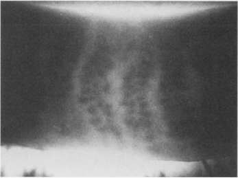
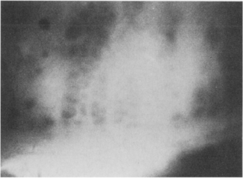
Figure 1. Top: Meibography of a normal eyelid. Bottom: Abnormal dilation of meibomian glands in a patient with meibomian gland dysfunction.
- Confocal microscopy
- Adjunctive ocular surface/tear film testing
- Tear film break-up time (TBUT) and Schirmer testing – used alongside meibography to correlate structural changes with tear film instability
- Lipid lay interferometry- device-based assessment of tear lipid layer thickness and dynamics as a functional correlate of meibum quality, now incorporated into MGD workup alongside meibography
- Demodex-specific testing
- Lash epilation and light microscopy (Gao, 2005)
- Collarette assessment in downgaze at slit-lamp (Trattler, 2022)
- Chalazia – diagnosis is clinical
- Biopsy indicated for atypical, recurrent or unilateral lesions to exclude masquerading etiologies (sebaceous gland carcinoma)
- Cultures are generally not helpful
- Yield is higher with frank purulence
- Staph epidermidis may be interpreted as normal flora even if clinically significant
- Culture for MRSA can support treatment, if oral antibiotics prescribed for acute signs of infection
Risk factors
Blepharitis
- Dry eye syndrome
- Dermatologic conditions: e.g., seborrheic dermatitis
- Rosacea
- Oral retinoid therapy
- Demodicosis
- Giant papillary conjunctivitis
Chalazia/Hordeola
- Rosacea
- Chronic posterior blepharitis (meibomian gland dysfunction)
- Demodex might be a risk factor (Yam 2014).
Differential diagnosis
Malignant tumors
- Sebaceous gland carcinoma
- Basal cell carcinoma
- Squamous cell carcinoma
- Merkel cell carcinoma (Rawlings 2007)
- Hemangioendothelioma (Al-Faky 2011)
- Plasmacytoma (Maheshwari 2009)
- Renal cell carcinoma (Tailor 2008)
- Pleural mesothelioma (Tsina 2006)
Benign tumors
- Pleomorphic adenoma (Ramlee 2007)
- Granular cell tumor (Scruggs 2014)
- Solitary neurofibroma (Shibata 2012)
- Plexiform neurofibroma (Tey 2006)
- Pilomatrixoma (Katowitz 2003)
- Keratoacanthoma
- Papilloma
- Ruptured epithelial inclusion cyst
Inflammatory disorders
- Sarcoidosis
- Atypical mycobacterial infection
- Wegener granulomatosis (Ismail 2007)
- Ruptured epithelial inclusion cyst
- Hyperimmunoglobulinemia E (Job) syndrome (Destefano 2004, Crama 2004)
Infectious
- Preseptal cellulitis
- Canaliculitis (Almaliotis 2013)
- Tuberculosis (Mittal 2013)
- Leishmaniasis (Rahimi 2009)
Other
- Retained soft contact lens (Agarwal 2013)
- Trichilemmal cyst (Meena 2012)
- Lacrimal gland duct stones (Kim 2014)
Patient management: treatment and follow-up
Natural history
- Blepharitis
- Chronic, with periods of exacerbation and remission
- Can begin in childhood, although typically onset is in middle age
- If severe, can cause eyelash loss, scarring of eyelids, trichiasis, secondary corneal scarring
- Chalazia
- Typically self-limiting, resolving in 1–2 weeks
- Chronic form may develop which can take many months to years to resolve.
- Eventually, virtually all chalazia resolve, even after years (Honda 2010).
Medical therapy
Blepharitis
- Eyelid hygiene
- Warm compresses
- Scrubs with water, dilute baby shampoo or commercially available eyelid wipes once or twice daily depending on severity
- Olenik study (Olenik 2013):
- Randomized double-blinded trial of baby-shampoo eyelid cleaning and preservative-free artificial tears with placebo or 1.5 gram of a composition (DHA, EPA, vitamins A, C, and E, tyrosine, cysteine, glutathione, zinc, copper, manganese, selenium, DPA)
- Measurements of staining, tear breakup time, Schirmer test, eyelid inflammation, ocular surface disease index (OSDI), meibomian gland expression
- After 3 months (61 patients), there was significant improvement of OSDI, TBUT, eyelid margin inflammation, meibomian gland expression, Schirmer test.
- Recent studies have questioned the efficacy of omega-3 supplementation for dry eye over placebo (DREAM study)
- For staphylococcal blepharitis treat with topical antibiotics, for example, erythromycin or bacitracin ophthalmic ointment
- Topical azithromycin ophthalmic solution 1%
- Has anti-inflammatory properties
- Studies have shown improvement for anterior and posterior blepharitis — open label studies (John 2008, Luchs 2008, Haque 2010, Opitz 2011).
- Dosing schedules are varied – 1 gtt BID x 2 days, then qday for 7–28 days (Opitz 2012)
- A multicenter, randomized study comparing Tobradex ST (4x/day x 14 days) to Azasite (2x/day x 12 days) for blepharitis favored Tobradex (Torkildsen 2011)
- Demodex-directed therapy
- First-line, FDA-approved: lotilaner ophthalmic solution 0.25% (Xdemvy, Tarsus Pharmaceuticals), approved July 2023; 1 drop BID for 6 weeks; GABA-gated chloride channel inhibitor causing mite paralysis and death (Gaddie 2023, Yeu 2023)
- DEPTH expert panel consensus (2023) provides treatment algorithm based on collarette severity and treatment response
- Tea tree oil/ terpinene-4-0l
- Previously first-line, now second-tier – a 2024 meta-analysis of 6 studies found unclear clinical efficacy and demonstrated in vitro toxicity to human meibomian gland epithelial cells
- Ivermectin
- Topical 1% cream can reduce mite density
- Oral at a dose of 200 mcg/kg once, repeated in 7 days – reduces demodex folliculorum in refractory blepharitis (Holzchuh, 2011)
- Systemic tetracyclines for chronic meibomitis/rosacea-associated disease
- Minocycline or doxycycline 50–100 mg BID, can taper to qday after the first month
- Slow-release formulation (50 mg qday) might be effective
- Alternatives include tetracycline 500 mg BID or azithromycin 250–500 mg, 1–3x/week; or 1g qweek x 3 weeks (caution in patients with cardiac conduction abnormalities)
- Risks: Photosensitization, gastrointestinal (GI) upset, azotemia, candidiasis
- Use is contraindicated in children and pregnant or nursing women.
- AO Ophthalmic Technology Assessment on oral antibiotics for meibomian gland-related OSD concluded there is no Level I evidence supporting oral doxycycline, minocycline, or azithromycin (Waldis 2016); 2024 review confirmed findings with better quality pooled data (Ben Ephraim 2024)
- Topical corticosteroids
- Reasonable for severe inflammatory flares, per current practice pattern guidance; not indicated for chronic maintenance given side-effect profile.
- Risk: Increased intraocular pressure, cataract, infection
- Devise-based therapy
- Eyelid thermal pulsation system (LipiFlow)
- Single 12-minute treatment improves OSDI more than warm compresses alone (Tao 2023)
- Intense pulsed light (IPL)
- Evidence with mechanistic rationale extending to Demodex mite eradication via chromophore-targeted thermal coagulation (Fineide 2024)
Meibomitis
- Literature assessment by the American Academy of Ophthalmology (and reinforced by recent metat-analysis) concluded that there is no level I evidence to support use of oral antibiotics including doxycycline, minocycline or azithromycin for meibomian gland related ocular surface disease (Wladis, 2016, Ben Ephraim 2024)
Chalazia/Hordeola
- Warm compresses and eyelid massage
- Eyelid hygiene/scrubs
- Topical antibiotics have limited evidence of efficacy even for the staphylococcal component — a large retrospective review found antibiotic use in only 36.5% of treated patients without clear efficacy signal and Cochrane review found topical antibiotics generally ineffective for acute internal hordeolum Systemic antibiotics are active against Staphylococcus aureus for accompanying preseptal cellulites.
- Systemic tetracyclines for treatment of chronic accompanying meibomitis, rosacea
- IPL chronic/recurrent chalazion and hordeolum, now with dedicated peer-reviewed trial data (Jiang 2023)
- Consider demodex screening in recurrent/multiple chalazion given strong association
- Topical steroid can be used to decrease inflammatory component of skin (although not demonstrated in any studies).
Surgery
Blepharitis
- Intraductal meibomian probing (that is, Maskin probe) (Maskin 2010)
- Office-based procedure using topical or injected local anesthesia, then progressive orifice/intraductal probing with a rigid stainless-steel probe
- Technique: start with a 2-mm probe (Figure 2) or hyfrecator tip (Wladis 2012); progress to 4-mm probe if resistance is encountered at the orifice or within the gland (fibrovascular tissue); a droplet of blood at the orifice after penetration is a normal finding, not a complication
- Improvement in OSDI symptom scores, though no objective improvement was seen on exam findings or meibum analysis in this small study, an important caveat for fellows (Wladis 2012)
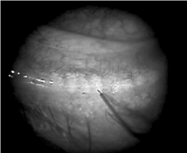
Figure 2. Maskin probe usage. Penetration through orifice with 2-mm probe. Note hemorrhage at orifice of adjacent gland.
- Can encounter resistance at meibomian gland orifice and within gland (fibrovascular tissue)
- Normal to have droplet of blood at orifice
- Then repeat with 4-mm probe
- Can be also done with hyfrecation tip (Figure 3) (Wladis 2012)
- Anatomic changes resulting in trichiasis or entropion might require surgical correction.
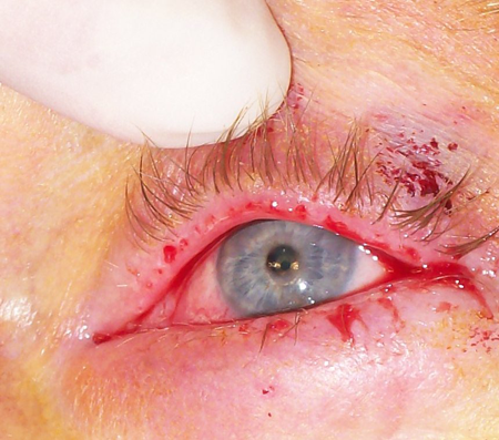
Figure 3. Meibomian probing. Image courtesy Edward J. Wladis, MD.
Chalazia/Hordeola
- Intralesional/perilesional corticosteroid injection
- Can be used for small marginal lesions or other surgically inconvenient lesions
- Multiple injections may be needed
- Can use 40 mg/mL or 10 mg/mL triamcinolone and inject 0.05 to 0.15 cc; dexamthasone 10mg/mL also used
- Complication profile per current AAO summary: skin depigmentation (especially in dark-skinned patients), subcutaneous fat atrophy, and rare but serious ocular penetration or embolization with vision loss.
- Surgical drainage via a transconjunctival or cutaneous route
- Meta-analysis found surgical curettage more successful than steroid injection for a single procedure, though the gap narrows when multiple procedures are permitted for either arm (Aycinena 2016)
- Technique
- Especially with small lesions, consider marking the skin prior to injection; otherwise, the location of the chalazion can be masked after infiltration.
- Consider using a pledget with 4% lidocaine (plain) between the globe and chalazion to help dull the pain of injection.
- Incision and curettage:
- Chalazion clamp is placed on the eyelid to isolate the chalazion, and the eyelid is everted, -a stab incision through the posterior tarsal plate is made with a #11 or 15 blade ( vertical or an “x;” some surgeons excise the flaps of the “x;” ), liquid and gelatinous material is expressed and curettage can be performed, granulomatous tissue and cyst wall excised
- Cutaneous excision
- Reserved for cases with skin-level changes (erythema, infiltration, thinning); caution regarding visible scarring in darker skin tones; tarsal plate component must still be addressed.
- Because the source of the chalazion is in the tarsal plate, this must also be addressed in addition to the skin.
- Trephination
- Through the conjunctival surface, a “punch” biopsy trephine can be used (Leachman).
- With the trephine (2–5 mm diameter), center it over the visible lesion, and then slowly rotate the trephine while applying pressure.
- Be careful to prevent penetration past the tarsal plate.
- Once through the full thickness of tarsus, use scissors and forceps to excise.
- Margin lesions
- Some suggest marginal curettage (Dubey).
- After placement of an appropriately sized clamp, the curette is placed into the chalazion and curetted.
- The remainder of the proximal chalazion is approached in the “standard” fashion.
- Care should be taken not to communicate the 2 areas to prevent notching.
- Combined excision and corticosteroid injection
- After excision is completed, some authors advocate for intratarsal injection of steroid.
- Best done with clamp in place to prevent embolization of steroid material
- Biopsy for recurrent or atypical lesions
- Caution for any atypical lesions in either appearance or history
- Concern for malignancy or other atypical lesions
Preventing and managing treatment complications
Pyogenic granuloma formation
- Occurs in the presence of an underlying chalazion/hordeolum
- After excision of pyogenic granuloma, also excise the chalazion.
Globe perforation
- Results in severe visual loss
- Perforation during excision — 2 cases (Shiramizu 2004)
- Perforation during injection — 1 case (Hosal 2003)
Eyelid margin scarring or notching
- Use care when removing tissue at or near the lid margin because there is a risk for eyelid scarring or notching.
- Can revise severe scar or notch with full-thickness pentagonal wedge resection
- Horizontal scarring of tarsal plate
- Some suggest prevention by making only vertical incisions along meibomian gland.
- Make an enlarged excision of involved tarsus to prevent vertical shortening of eyelid.
- Repeated episodes or surgical treatments can lead to posterior lamellar scarring and misdirected eyelashes or eyelash loss.
Damage to surrounding meibomian glands
- Prevent by making vertical cuts through tarsal conjunctival surface.
Damage to punctum or canaliculus
- Use caution when excising peripunctal or pericanalicular chalazia.
- If punctum or canaliculus is violated, repair with silicone stent intubation.
References and additional resources
- American Academy of Ophthalmology Cornea/External Disease Panel. Preferred Practice Pattern Guidelines. Blepharitis. 2013. Available at aao.org/ppp.
- Basic and Clinical Science Course. Section 4: Ophthalmic Pathology and Intraocular Tumors; Section 6: Pediatric Ophthalmology and Strabismus; Section 7: Orbit, Eyelids, and Lacrimal System; Section 8: External Disease and Cornea, 2013-2014. San Francisco: American Academy of Ophthalmology.
- Focal Points: Diagnosis and Treatment of Chronic Blepharitis, Module 10, 1989. San Francisco: American Academy of Ophthalmology.
- Monograph 8, Surgery of the Eyelids, Lacrimal System, & Orbit, 2nd edition, 2011. San Francisco: American Academy of Ophthalmology.
- Agarwal PK, Ahmed TY, Diaper CJ. Retained soft contact lens masquerading as a chalazion: a case report. Indian J Ophthalmol. 2013;61(2):80-81.
- Al-Faky YH, Al Malki S, Raddaoui E. Hemangioendothelioma of the eyelid can mimic chalazion. Oman J Ophthalmol. 2011;4(3):142-143.
- Almaliotis D, Nakos E, Siempis T, et al. A para-canalicular abscess resembling an inflamed chalazion. Case Rep Ophthalmol Med. 2013;618367.
- Arita R, Minoura I, Morishige N, et al: Development of definitive and reliable grading scales for meibomian gland dysfunction. Am J Ophthalmol 2016; 169:125-137.
- Aronowicz JD, Shine WE, Oral D, et al. Short term oral minocycline treatment of meibomianitis. Br J Ophthalmol 2006;90:856–60.
- Aycinena AR, Achiron A, Paul M, Burgansky-Eliash Z. "Incision and Curettage Versus Steroid Injection for the Treatment of Chalazia: A Meta-Analysis." Ophthalmic Plast Reconstr Surg. 2016;32(3):220-224
- Ben Simon GJ, Rosen N, Rosner M, Spierer A. Intralesional triamcinolone acetonide injection versus incision and curettage for primary chalazia: a prospective, randomized study. Am J Ophthalmol. 2011;151(4):714-718.
- Ben Ephraim Noyman D, Chan CC, Mimouni M, et al. "Systemic antibiotic treatment for meibomian gland dysfunction: a systematic review and meta-analysis." Acta Ophthalmol. 2024;102(1):e1-e10
- Bowman RW, Dougherty JM, McCulley JP. "Chronic blepharitis and dry eyes." Int Ophthalmol Clin. 1987;27(1):27-35
- Cac NN, Messingham MJ, Sniezek PJ, Walling HW. Stevens-Johnson syndrome induced by doxycycline. Cutis 2007; 79:119-122.
- Cohen BZ, Tripathy RC. Eyelid depigmentation after intralesional injection of a fluorinated corticosteroid for chalazion. Am J Ophthalmol. 1979;88(2):269-270.
- Crama N, Toolens AM, van der Meer JW, Cruysberg JR. Giant chalazia in the hyperimmunoglobulinemia E (hyper-IgE) syndrome. Eur J Ophthalmol. 2004;14(3):258-260.
- Destefano JJ, Kodsi SR, Primack JD. Recurrent Staphylococcus aureus chalazia in hyperimmunoglobulinemia E (Job’s) syndrome. Am J Ophthalmol. 2004;138(6):1057-1058.
- Dougherty JM, McCulley JP, Silvany RE, Meyer DR. The role of tetracycline in chronic blepharitis: inhibition of lipase production in staphylococci. Invest Ophthalmol Vis Sci. 1991;32:2970-2975.
- Driver PJ, Lemp MA. Meibomian gland dysfunction. Surv Ophthalmol. 1996;40(5):343-347.
- Dubey R, Wang LW, Figueira EC, et al. Management of marginal chalazia: a surgical approach. Br J Ophthalmol. 2011;95:590-597.
- Duncan & Freitag, Rev Ophthalmol. 2022; AAO Blepharitis PPP, Ophthalmology. 2019
- Edwards RS. Ophthalmic emergencies in a district general hospital casualty department. Br J Ophthalmol. 1987;71:938-942.
- Favier C. Demodex clusters. Ophthalmology 2017; 124:1474.
- Fineide F, et al. Acta Ophthalmol. 2024;102:e414-e442
- Frucht-Pery J, Sagi E, Hemo I, Ever-Hadani P. Efficacy of doxycycline and tetracycline in ocular rosacea. Am J Ophthalmol. 1993;116:88-92.
- Gao YY, Di Pascuale MA, Li W, et al. High prevalence of Demodex in eyelashes with cylindrical dandruff. Invest Ophthalmol Vis Sci 2005;46(9):3089–3094.
- Goawalla A, Lee V. A prospective randomized treatment study comparing three treatment options for chalazia: triamcinolone acetonide injections, incision and curettage and treatment with hot compresses. Clin Experiment Ophthalmol. 2007;35(8):706-712.
- Greene JB, Jeng BH, Fintelmann RE, et al: Oral azithromycin for the treatment of meibomitis. JAMA Ophthalmol 2014; 132:121-122.
- Greiner JV. A single LipiFlow thermal pulsation system treatment improves Meibomian gland function and reduces dry eye symptoms for 9 months. Curr Eye Res. 2012;37(4):272‑278.
- Haque RM, Torkildsen GL, Brubaker K, et al. Multicenter open-label study evaluating the efficacy of azithromycin ophthalmic solution 1% on the signs and symptoms of subjects with blepharitis. 2010;29:871‑877.
- Hassanzadeh S, Varmaghani M, Zarei-Ghanavati S, Heravian Shandiz J, Azimi Khorasani A. Ocul Immunol Inflamm. 2021;29(1):66-75
- HolzchuhFG, Hida RY, Moscovici BK, et al: Clinical treatment of ocular demodex folliculorum by systemic ivermectin. Am J Ophthalmol 2011; 151:1030-1034.
- Hom MM, Martinson JR, Knapp LL, et al. Prevalance of Meibomian gland dysfunction. Optom Vis Sci. 1990;67:710‑712.
- Honda M, Honda K. Spontaneous resolution of chalazion after 3 to 5 years. Eye Contact Lens. 2010;36(4):230‑232.
- Hosal BM, Zilelioglu G. Ocular complication of intralesional corticosteroid injection of a chalazion. Eur J Ophthalmol. 2003;13(9‑10):798‑799.
- Igami TZ, Holzchuh R, Osaki TH, et al. Oral azithromycin for treatment of posterior blepharitis. Cornea 2011;30:1145–9.
- Ismail AR, Theaker JM, Manners RM. Wegener’s granulomatosis masquerading as upper lid chalazion. Eye (Lond). 2007;21(6):883‑884.
- IovienoA, LambiaseA, MiceraA, etal. In vivo characterization of doxycycline effects on tear metalloproteinases in patients with chronic blepharitis. Eur J Ophthalmol 2009;19:708–16.
- Jacobs PM, Thaller VT, Wong D. Intralesional corticosteroid therapy of chalazia: a comparison with incision and curettage. Br J Ophthalmol. 1984;68:836‑837.
- Jiang J, Yang Z, Du F, et al. "Therapeutic effect of intense pulsed light on different types of chalazion in children." Sci Rep. 2024;14:3645; Yang K, Wen Y, Zhu L, et al. "The Effect of Intense Pulsed Light Treatment for Chronic Hordeolum." Biomed Environ Sci. 2023;36:1005-1014
- John T. Use of azithromycin ophthalmic solution in the treatment of chronic mixed anterior blepharitis. Ann Ophthalmol. 2008;40(2):68‑74.
- Katowitz WR, Shields CL, Shields JA, et al. Pilomatrixoma of the eyelid simulating a chalazion. J Pediatr Ophthalmol Strabismus. 2003;40(4):247‑248.
- Kheirkhah A, Blanco G, Casas V, Tseng SC. Fluorescein dye improves microscopic evaluation and counting of Demodex in blepharitis with cylindrical dandruff. Cornea 2007;26(6): 697–700.
- Kim SC, Lee K, Lee SU. Lacrimal gland duct stones: misdiagnosied as chalazion in 3 cases. Can J Ophthalmol. 2014;49(1):102‑105.
- Lane SS, DuBiner HB, Epstein RJ, et al. A new system, the LipiFlow, for the treatment of Meibomian gland dysfunction. 2012;31:396‑404.
- Lau B, Mutyala D, Dhaliwal D. A case report of doxycycline-induced Stevens-Johnson syndrome. Cornea 2011; 30:595-597.
- Leachman JW, Boynton JR, Levin DB. Chalazion management by tarsus trephination. Ophthalmic Surg. 1978;9(1):89‑90.
- Lemp MA, Nichols KK. Blepharitis in the United States: a survey based perspective on prevalence and treatment. Ocul Surf. 2009;7(Suppl 2):S1‑S14.
- Liang L, Ding X, Tseng SCG. High prevalence of demodex brevis infestation in chalazia. Am J Ophthalmology 2014; 342-348.
- Lindsley K, Matsumura S, Hatef E, Akpek EK. Interventions for chronic blepharitis. Cochrane Database Syst Rev. 2012;5:CD005556.
- Luang J, Guo MX, Xiang DM, et al. BMC Ophthalmol. 2022;22:124
- Luchs J. Efficacy of topical azithromycin ophthalmic solution 1% in the treatment of posterior blepharitis. Adv Ther. 2008;25(9):858‑870.
- Maheshwari R, Maheshwari S. Extramedullary plasmacytoma masquerading as chalazion. 2009;28(2‑3):191‑193.
- Maskin SL. Intraductal Meibomian gland probing relieves symptoms of obstructive Meibomian gland dysfunction. 2010;29(10):1145‑1152.
- Mathers WD, Shields WJ, Sachdev MS, et al. Meibomian gland dysfunction in chronic blepharitis. 1991;10:277‑285.
- McCulley JP, Dougherty JM, Deneau DG. Classification of chronic blepharitis. Ophthalmology. 1982;89(10):1173‑1180.
- McCulley JP, Dougherty HM. Blepharitis associated with acne rosacea and seborrheic dermatitis. Int Ophthalmol Clin. 1985;25(1):159‑172.
- Meena M, Mittal R, Saha D. Trichilemmal cyst of the eyelid: masquerading as recurrent chalazion. Case Rep Ophthalmol Med. 2012;261414.
- Meibomius H. De Vasis Palpebrarum Novis Epistola Muller, Helmstadt, 1666.
- Mittal R, Tripathy D, Sharma, Balne PK. Tuberculosis of eyelid presenting as a chalazion. Ophthalmology. 2013;120(5):e1‑4.
- National Disease and Therapeutics Index (NDTI), IMS America, Dec 1982.
- Nelson JD, Shimazaki J, Benitez-del-Castillo JM, et al. Invest Ophthalmol Vis Sci. 2011;52(4):1930-1937
- Nelson JD, et al. IOVS. 2011;52(4):1930-1937; Sheppard JD, Nichols KK. Ophthalmol Ther. 2023;12(3):1397-1418)
- Nemet AY, Vinker S, Kaiserman I. Associated morbidity of blepharitis. Ophthalmology. 2011;118:1062‑1068.
- Nichols JJ, Berntsen DA, Mitchell GL, et al. An assessment of grading scales for meibography images. 2005;24:382‑388.
- Olenik A, Jimenez-Alfaro I, Alejandre-Alba N, Mahillo-Fernandez I. A randomized, double-masked study to evaluate the effect of omega‑3 fatty acids supplementation in Meibomian gland dysfunction. Clin Interventions Aging. 2013;8:1133‑1138.
- Opitz DL, Tyler KF. Efficacy of azithromycin ophthalmic solution for treatment of ocular surface disease from posterior blepharitis. Clin Ep Optom. 2011;94(2):200‑206.
- Opitz DL, Harthan JS. Review of azithromycin ophthalmic 1% solution (AzaSite) for the treatment of ocular infections. Ophthalmol and Eye Dis. 2012;4:1‑14.
- Ozdal PC, Codere F, Callejo S, et al. Accuracy of the clinical diagnosis of chalazion. Eye (Lond). 2004;18(2):135‑138.
- Papas A, Singh M. The effect of a unique omega‑3 supplement on dry mouth and dry eye in Sjogrens patients. ARVO Annual Meeting. 2007.
- Peralejo B, Beltriani V, Bielory L. Dermatologic and allergic conditions of the eyelid. Immunol Allergy Clin North Am. 2008;28:137‑168.
- Pinheiro MH Jr, Dos Santos PM, Dos Santos RC, et al. Oral flaxseed oil (Linum usitatissimum) in the treatment for dry-eye Sjogren’s syndrome patients. Arq Bras Oftalmol. 2007;70:649‑655.
- Quarterman MJ, Johnson DW, Abele DC, et al. Ocular rosacea. Signs, symptoms, and tear studies before and after treatment with doxycycline. Arch Dermatol 1997;133:49–54.
- Rahimi M, Moinfar N, Ashrafi A. Eyelid leishmaniasis masquerading as chalazia. Eye (Lond). 2009;23(3):737.
- Raju VK, Raju LV, Kheirkah A. Demodex blepharitis. Ophthalmology 2012; 119-200.
- Ramlee N, Ramli N, Tajudin LS. Pleomorphic adenoma in the palpebral lobe of the lacrimal gland misdiagnosed as chalazion. 2007;26(2):137‑139.
- Rawlings NG, Brownstein S, Jordan DR. Merkel cell carcinoma masquerading as a chalazion. Can J Ophthalmol. 2007;42(3):469‑470.
- Robin JB, Jester JV, Nobe J, et al. In vivo transillumination biomicroscopy and photography of Meibomian gland dysfunction: a clinical study. Ophthalmology. 1985;82:1423‑1426.
- Scruggs RT, Black EH. Granular cell tumor masquerading as a chalazion: a case report. Ophthal Plast Reconstr Surg. 2015;31(1):e6‑8.
- Sherman RS, Hogan MJ. Radiation therapy in diseases of the eye. California Medicine. 1954;80(2):83‑90.
- Shibata N, Kitagawa K, Noda M, Sasaki H. Solitary neurofibroma without neurofibromatosis in the superior tarsal plate simulating a chalazion. Graefes Arch Clin Exp Ophthalmol. 2012;250(2):309‑310.
- Shine WE, McCulley JP, Pandya AG. Minocycline effect on meibomian gland lipids in meibomianitis patients. Exp Eye Res. 2003;76:417‑420.
- Shiramizu KM, Krieger AE, McCannel CA. Severe visual loss caused by ocular performation during chalazion removal. Am J Ophthalmol. 2004;137(1):204‑205.
- Sobolewska B, Doycheva D, Deuter C, et al. Treatment of ocular rosacea with once-daily low-dose doxycycline. 2014;33(3):257‑260.
- Tailor R, Inkster C, Hanson I, et al. Metastatic renal cell carcinoma presenting as a chalazion. Eye (Lond). 2008;21(4):564‑565.
- Tao JP, Shen JF, Aakalu VK, et al. "Thermal Pulsation in the Management of Meibomian Gland Dysfunction and Dry Eye: A Report by the American Academy of Ophthalmology." Ophthalmology. 2023;130(12):1336-1341
- Tey A, Kearns PP, Barr AD. Plexiform neurofibroma masquerading as a persistent chalazion – a case report. Eye (Lond) 2006;20(8):946‑948.
- Thomas EL, Laborde RP. Retinal and choroidal vascular occlusion following intralesional corticosteroid injection of a chalazion. Ophthalmology. 1986;93:405‑407.
- Torkildsen GL, Cockrum P, Meier E, et al. Evaluation of clinical efficacy and safety of tobramycin/dexamethasone ophthalmic suspension 0.3%/0.05% compared to azithromycin ophthalmic solution 1% in the treatment of moderate to severe acute blepharitis/blepharoconjunctivitis. Curr Med Res Opin. 2011;27(1):171‑178.
- Trattler W, Karpecki P, Rapoport Y, et al. Clin Ophthalmol. 2022;16:1153-1164
- Tsina EK, Papioannou D, Matsouka FO, Kosmidis PA. Metastatic pleural mesothelioma initially masquerading as chalazion. Br J Ophthalmol. 2006;90(7):921‑922.
- US Food and Drug Administration. FDA Drug Stafety Communication. Azithromycin (Zithromax or Zmax) and the risk of potentially fatal heart rhythms. Available at fda.gov/drugs/drugsafety/ucm341822.htm.
- Winn BJ, Liao YJ, Horton JC. Intracranial pressure returns to normal about a month after stopping tetracycline antibiotics. Arch Ophthalmol 2007;125(8):1137‑1138.
- Wladis EJ. Intraductal Meibomian gland probing in the management of ocular rosacea. Ophthal Plast Reconstr Surg. 2012;28:416‑418.
- Wladis EJ, Bradley EA, Bilyk JR, el al: Oral antibiotics for meibomian gland-related ocular surface disease: A report by the American Academy of Ophthalmology. Ophthalmology 2016; 123:492-496.
- Wojtowicz JC, Butovich I, Uchiyama E, et al. Pilot, prospective, randomized, double-masked, placebo-controlled clinical trial of an omega‑3 supplement for dry eye. 2011;30:308‑314.
- Yagci A, Palamar M, Egrilmez S, et al. Anterior segment ischemia and retinochoroidal vascular occlusion after intralesional steroid injection. Ophthal Plast Reconstr Surg. 2008;24(1):55‑57.
- Yam JC, Tang BS, Chang TM, Cheng AC. Ocular demodicidosis as a risk factor of adult recurrent chalazion. Eur J Ophthalmol. 2014;24(2):159‑163.
- Yoo SE, Lee DC, Chang MH. The effect of low-dose doxy- cycline therapy in chronic meibomian gland dysfunction. Korean J Ophthalmol 2005;19:258–63.
Financial disclosures
Reviewers
Kate Lane: None