
Eyelid Degenerative and Inflammatory Disorders
Updated July 2026
Acrochordon (skin tag)
Approach to diagnosis
- Etiology: thought to be caused by skin rubbing
- Appearance in childhood can be associated with basal cell nevus syndrome.
- Increased incidence with age, no gender/race predilection
- Characteristic appearance: pedunculated, fleshy, skin-colored mass
- Benign, slow growing, usually asymptomatic, sometimes causes irritation
- Most often seen in neck, axilla, and inguinal region
- Definitive diagnosis by histopathology
Risk factors
- Obesity
- Diabetes
Differential diagnosis
- Squamous papilloma
- Verrucca vulgaris
- Amelanotic nevus
Clinical management
- Observation
- Surgical excision
- Cryotherapy
Complications of treatment
Rare; recurrence uncommon
Squamous papilloma
Approach to diagnosis
- Hyperplasia of squamous epithelium
- Characteristic appearance: frond-like, papular lesion, often pedunculated
- Slow growing, usually asymptomatic
- Definitive diagnosis by histopathology
- Fibrovascular core covered by acanthotic epithelium, hyperkeratosis
Risk factors
- Increased incidence with age, no gender/race predilection
- Most common benign eyelid lesion
Differential diagnosis
- Verrucca vulgaris
- Amelanotic nevus
- Seborrheic keratosis
- Basal cell carcinoma
Clinical management
- Observation
- Surgical excision
- Laser ablation (CO2, argon)
Complications of treatment
Rare; recurrence uncommon
Seborrheic keratosis (SK)
Approach to diagnosis
- Unknown etiology; arises from follicular infundibulum, involves aggregation of immature epidermal keratinocytes
- Increased incidence with age, no gender/race predilection
- Clinical features: elevated, sometimes pedunculated, rough keratotic surface with variable pigmentation, greasy “stuck-on” appearance
- Slow growing, usually asymptomatic; sometimes causes irritation
- Commonly seen on torso and face
- Clinical variants:
- Dermatosis papulosa nigra: Multiple deeply pigmented facial SKs in Africans
- Inverted follicular keratosis: Irritated variant of SK, rapid growth typical
- Intraepidermal horn cysts: Thick protuberance of keratin from SK
- Sudden appearance or growth of multiple SKs can herald internal malignancies such as adenocarcinoma of GI or breast (Leser-Trelat sign)
- Definitive diagnosis by histopathology: Hyperkeratosis, papillomatosis, acanthosis
Risk factors
- Family history
- Sun exposure
- Age
Differential diagnosis
- Squamous papilloma (pigmented)
- Nevus
- Melanoma
- Pigmented basal cell carcinoma
Clinical management
- Observation
- Surgical excision
- Cryotherapy
Complications of treatment
Rare; recurrence uncommon
Pseudoepitheliomatous hyperplasia (PEH)
Approach to diagnosis
- Pseudoneoplastic proliferation of epidermis and adnexal epithelium
- Can be idiopathic or associated with numerous types of infection (classically mycotic or mycobacterial), inflammation, trauma, or neoplasms (for example, basal cell carcinoma)
- Epidemiology: increased incidence with age
- History:
- Can develop rapidly over weeks
- Can gradually regress if left untreated for several months
- Clinical features:
- Nodular, irregular, crusty lesion, sometimes ulcerative
- Often resembles cutaneous malignancy
- Variant: keratoacanthoma (see Squamous Cell Carcinoma)
- Diagnosis established by histopathology:
- Irregular invasion of dermis by jagged epidermal masses, horn cysts, mitotic figures, nonspecific inflammation
- Can be difficult to distinguish from squamous cell carcinoma
- Inciting organism can be seen if present
Risk factors
Presence of inciting condition
Differential diagnosis
- Squamous cell carcinoma
- Basal cell carcinoma
Patient management
- Observation (can spontaneously regress)
- Medical therapy
- Treatment of underlying condition if identified
- Surgical excision
- If diagnostic uncertainty exists, consider Mohs excision or frozen section control
Complications of treatment
Scarring, eyelid deformity
Sebaceous hyperplasia
Approach to diagnosis
- Etiology unknown
- Epidemiology:
- Increased incidence with age, most common over 40–50
- Rare familial forms exist
- History: gradual onset, chronic course
- Clinical features:
- One or more focal tan-yellow papules or diffuse thickening of eyelids
- Similar to sebaceous adenoma
- Associated with Muir-Torre syndrome (MTS):
- Sebaceous neoplasms or keratoacanthoma
- Internal malignancy (for example, colorectal carcinoma)
- Presence of sebaceous hyperplasia does not necessitate screening for MTS
- Diagnosis established by histopathology: Well-differentiated, mature sebaceous lobules
Risk factors
- Muir-Torre syndrome
- Family history
Patient management
- Observation
- Trichloroacetic acid (small, diffuse lesions)
- Surgical options:
- Excision
- Electrodessication, cautery (small lesions)
Xanthelasma
Approach to diagnosis
- Etiology: idiopathic, sometimes associated with familial hyperlipidemia
- A large case-control study (n=35,452) found no significant association between xanthelasma and dyslipidemia or cardiovascular disease when rigorously matched ( Lustig-Barzelay, 2025)
- Epidemiology:
- 1–3% incidence in general population, generally between 35 and 55
- Affects women twice as often as men
- History: gradual onset, usually static, but can fluctuate in size/number
- Clinical features:
- Flat or mildly elevated, yellow, subepidermal placoid lesions
- Usually bilateral, can involve all 4 lids, medial location common
- Diagnosis usually clinical:
- Atypical appearance should raise suspicion for xanthogranulomatous disease (see below)
- Histopathology: infiltration of superficial reticular dermis foamy histiocytes, Touton giant cells sometimes present
Risk factors
Hyperlipidemia
Differential diagnosis
- Xanthoma
- Xanthogranuloma
Patient management (Yee, 2024)
- Observation
- Treatment of hyperlipidemia if present:
- Normalization of lipids can lead to regression
- Trichloroacetic or bichloroacetic acid
- Laser ablation (CO2, argon, erbium-YAG, pulsed dye), electrosurgery, cryotherapy, plasma and RF-based ablation
- Intralesional injection- bleomycin, pingyangmycin, heparin sodium
- Surgical excision:
- Typically full-thickness, but subepithelial excision if lipid infiltration extends beneath the surface
- Recurrence common regardless of treatment modality:
- Highest incidence of recurrence within first year
Complications of treatment
Cicatricial ectropion/lagophthalmos
Disease-related complications
Cardiovascular risk related to hyperlipidemia
Atopic dermatitis
Approach to diagnosis
- Unknown etiology; cell-mediated immune defects
- TH2-cell activation with increased synthesis of IL-4 and IgE
- Epidemiology:
- Manifests in infancy or childhood
- 2% of population affected by atopic dermatitis
- Characteristic triad:
- Eczematous dermatitis
- Allergic rhinitis
- Asthma
- Clinical features:
- Eyelid findings: eczema, pruritis, edema, violaceous or brown discoloration (“allergic shiner”), lichenification, periocular milia
- Dennie-Morgan fold: skin crease from medial canthus to lower lid
- Secondary staphylococcal infection can occur.
- Ocular findings: atopic keratoconjunctivitis, conjunctival papillae, Horner-Trantas dots, inferior pannus, symblepharon
- Keratoconus: 1.5–6.7% incidence in atopic dermatitis
- Skin hypersensitivity to numerous stimuli/agents
- Establishing the diagnosis
- History and physical exam
- Elevated serum IgE
Differential diagnosis
- Other forms of dermatitis: contact, seborrheic, lichen simplex chronicus
- Blepharochalasis syndrome
Risk factors
Family history of atopy
Patient management
- Natural history
- Infantile stage: eczematous dermatitis affecting extensor surfaces (tops of feet, back of hands, knees, face)
- Childhood phase: flexural sites (popliteal/antecubital fossae)
- Periodic exacerbations and remissions
- Medical therapy
- Break the “itch-scratch” cycle: cool moist compresses, bland ointments, antihistamines
- Acute flares: topical corticosteroids (5–10 days)
- Keratoconjuctivitis: topical antihistamines, mast-cell-stabilizers, corticosteroids, or NSAIDs
- Secondary staphylococcal infection: topical antibiotics
- Topical calcineurin inhibitors Pimecrolimus and Tacrolimus are both indicated for the treatment of atopic dermatitis.
- Safe and effective for sensitive areas, particularly given the low risk of causing atrophy.
- Negligible systemic levels after topical application.
- Boxed warning in US due to theoretical risk of lymphoma and nonmelanoma skin cancers, despite ample evidence suggesting no increased risk with topical use.
- Dupilumab (IL-4/IL-13 receptor antagonist) and topical/systemic JAK inhibitors (e.g., ruxolitinib cream, upadacitinib) have become standard escalation options for moderate-to-severe atopic dermatitis beyond topical calcineurin inhibitors, including periocular disease
- Surgery: not indicated
- Co-management with dermatology recommended
Complications of treatment
- Steroids: skin atrophy, glaucoma, cataract, susceptibility to infection
- Prevention by using short courses of treatment and avoiding long-term use
Contact dermatitis
Approach to diagnosis
- Etiology: exposure to offending agent
- Cosmetics, cleaning products, preservatives, metals, ophthalmic medications, and others
- Can be irritant or allergic (type IV hypersensitivity)
- Epidemiology: female predominance
- Clinical features:
- Acute: edema, erythema, pruritis, vesicles, weeping
- Chronic: erythema, lichenification, hyperpigmentation
- Patch testing with suspected offending agent
Differential diagnosis
- Other forms of dermatitis: atopic, seborrheic, lichen simplex chronicus
- Blepharochalasis syndrome
Patient management
- Remove offending agent
- Medical therapy
- Cool compresses, oral antihistamines
- Topical corticosteroid ointment
- Low-potency (hydrocortisone 1% cream/ointment, lotemax ointment)
- Topical dexamethasone ophthalmic suspension for severe/refractory cases
- Topical calcineurin inhibitors (tacorimus, pimecrolimus)
- Widely used off-label for allergic and irritant contact dermatitis (Approved for atopic dermatitis)
- Topical PDE-4 inhibitor (crisaborole) and topical JAK inhibitor (ruxolitinib 1.5% cream) are now included among alternatives to corticosteroids/calcineurin inhibitors for eyelid dermatitis
- Oral corticosteroids can be considered for acute cases
Complications of treatment and their prevention
- Steroids: skin atrophy, glaucoma, cataract, susceptibility to infection, topical steroid withdrawal (erythematous, edematous, burning plaques on steroid discontinuation)
- Prevention by using short courses of treatment and avoiding long-term use
Seborrheic dermatitis
Approach to diagnosis
- Etiology unknown; associated with increased sebaceous gland number and function
- Yeast (Pityrosporon ovale) thought to play a role
- Epidemiology:
- Infancy (“cradle cap”)
- Puberty through adulthood
- Male predilection
- Affects 2–5% of population
- History: gradual onset, worse during winter
- Clinical features:
- Greasy scaling, patchy erythema involving scalp, central face, lids, brow, ears, presternal skin, axilla, inguinal area
- Blepharitis, madarosis, keratoconjunctivitis
- Histopathology:
- Focal parakeratosis
- Pyknotic neutrophils
- Acanthosis
- Spongiosis
- Nonspecific inflammation of dermis
Risk factors
- Parkinson’s disease
- Epilepsy
- Multiple sclerosis
- AIDS
Differential diagnosis
- Psoriasis
- Impetigo
- Dermatophytosis
- Lichen simplex chronicus
- Discoid lupus
Patient management
- Natural history: chronic course with exacerbations and remissions
- Medical therapy:
- Anti-dandruff shampoos
- Ketoconazole shampoo 2% to face during shower or cream
- Corticosteroid cream
- Lid scrubs, topical antibiotics
- Radiation therapy: UV radiation helpful (condition improves in summer, flares in fall)
- Comanagement with dermatology recommended
Complications of treatment
Steroids:
- Skin atrophy
- Glaucoma
- Cataract
- Susceptibility to infection
Lamellar ichthyosis
Approach to diagnosis
- One phenotype within broader category of autosomal recessive congenital ichthyosis (ARCI)
- Etiology: genetic, usually autosomal recessive
- Identified mutations on chromosomes 14q11 and 2q33-35
- Abnormal keratin formation
- Subtype of ichthyosiform dermatoses which also include:
- Dominant ichthyosis vulgaris
- X-linked ichthyosis
- Epidermolytic hyperkeratosis
- Epidemiology
- Incidence 1:250,000–300,000
- History
- Disease evident at birth
- “Collodion baby”: encasement in transparent, parchment-like membrane that spontaneously sheds within 2 weeks
- Large scales develop after membrane sheds
- Clinical features
- Cicatricial ectropion and lagophthalmos (Figure 1A)
- Generalized fish-like scales over entire body (Figure 1B)
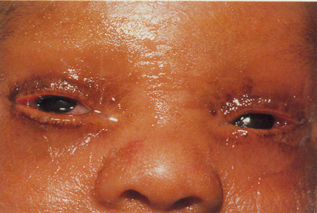
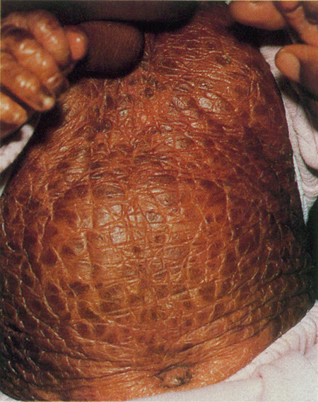
Figure 1. Two-week-old old infant with lamellar ichthyosis. A. Cicatricial lagophthalmos and ectropion of all four lids. B. Generalized scaling as demonstrated on abdomen (Oestreicher 1990).
- Corneal scarring: might not be completely attributable to exposure (Cruz 2000)
- Testing and evaluation for establishing the diagnosis
- Physical findings
- Skin biopsy
- Genetic testing
Risk factors
Family history
Differential diagnosis
Limited
Patient management (Mazereeuw-Hautier, 2025)
- Natural history: chronic and unremitting
- Medical therapy
- Skin moisturizers/emollients and keratolytics
- Less severe in warm, humid climates
- Vitamin D analogues (topical and systemic): emerging support
- Oral retinoids (acitretin) are used for severe ectropion and scaling
- JAK inhibitors may be emerging as targeted therapy
- Gene therapy- pre-clinical development, may result in disease-modifying treatment rather than treating symptoms
- Surgery: anterior lamellar grafts for cicatricial ectropion
- Donor sites for full-thickness skin grafts limited due to generalized involvement
- Penile foreskin (Uthoff 1994)
- Maternal allograft (Das 2010)
- Oral mucous membrane graft (Nayak 2011)
- Human engineered skin (Culican 2002)
- Inverting sutures (with systemic retinoids) for ectropion
- Advancement flaps
- Non-surgical options
- Hyaluronic acid filler injection for eyelid retractions/ectropion
- Scleral/PROSE lens, moisture goggles, amniotic membrane
Disease-related complications
Disease-related complications are syndrome-related: Refsum syndrome, Gaucher syndrome.
Dermatomyositis
Approach to diagnosis
- Classifications: antibody-defined phenotype model, dividing into 8 groups: adult DM, juvenile DM, amyopathic DM, cancer-associated DM, polymyositis, immune-mediated necrotizing myopathy, inclusion body myositis, and overlap myositis
- Myositis-specific antibodies now drive both prognosis and treatment selection
- Rather than a single autoantibody panel used mainly for diagnosis, specific antibodies now define distinct clinical phenotypes with different treatment algorithms
- The precise underlying autoimmune trigger remains incompletely understood
- Etiology: tyle I interferon-driven autoimmune vasculopathy
- Genetic susceptibility and environmental trigger model (viral infection)
- Auto-antibody-defined mechanism: specific antibody subtypes have been identified
- Environmental/infectious triggers
- Viral (coxsackievirus, parvovirus, HTLV-1, HIV, COVID-19)
- Drug-induced (for example, atorvastatin, phenytoin)
- Paraneoplastic: ovarian or gastric carcinoma, lymphoma, lung cancer, and others
- Epidemiology
- Can present in children (5–15 years) or middle-aged adults
- Female preponderance
- History: variable presentation, rash usually appears early
- Clinical features
- Periobital
- Bilateral heliotrope rash on upper lids or malar region (Figure 2)
- Periobital
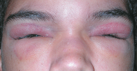
Figure 2. Heliotrope rash and edema of both upper eyelids in 16-year-old male with juvenile dermatomyositis (Taban 2006).
- Diffuse purple discoloration or scaly erythema — variant: supraciliary “purple line”
- Periorbital edema
- Pox-like scars (late stage)
- Combination of periorbital heliotrope erythema with eyelid edema (PHEEE) has been proposed as a specific clinical marker for paraneoplastic dermatomyositis, particularly in patients with anti-TIF1-γ or anti-NXP-2 autoantibodies (Aliu, 2024)
- Anterior and posterior segment findings of vasculopathy (episcleritis, iritis, cotton wool spots, vision loss)
- Cutaneous:
- Calcinosis cutis, a hallmark of chronic disease (mostly in children)
- Mechanic’s hand: fissured, scaly, hyperkeratotic, hyperpigmented hands
- Gottron’s papules (pathognomonic): scaly erythematous plaques over knuckles, elbows, and knees
- “Shawl” pattern rash over shoulders/arms/upper back/anterior neck/chest
- Gastrointestinal: Dysphagia, proximal muscle weakness
- Pulmonary: interstitial lung disease in 1/3 of patients; risk of morbity/mortality if present
- Arthritis, arthralgias- in 50% of patients, involving small joints
- Cardiac conduction defects
- Testing and evaluation for establishing the diagnosis
- 2017 EULAR/ACR Classification Criteria (EULAR/ACR Classification Criteria paper)calculates a probability score for idiopathic inflammatory myopathy (IIM) that classifies patients into possible/probable/definite categories rather than a binary yes/no
- If a pathognomonic DM skin rash (heliotrope, Gottron papules/sign) is present, the criteria can classify DM without requiring muscle biopsy; in the absence of a classic rash, biopsy is required for diagnosis
- Labs: creatine kinase (CK), aldolase, lactate dehydrogenase (LDH), liver enzymes, ANA, Myositis-specific and myositis-associated autoantibody (MSA/MAA) panel
- Electromyography – can guide muscle biopsy site selection
- MRI- evidence of active myositis within 6 months- supporting evidence
- Muscle biopsy- required when pathognomonic skin findings absent
- Mixed B- and T-cell perivascular infiltrate
- Perifascicular muscle fiber atrophy
- Skin biopsy- nonspecific findings overlapping with lupus erythematosus
- Systemic evaluation:
- pulmonary (CT chest, pulmonary function tests)
- malignancy screening- based on antibody profile
Risk factors
- Viral infection, possibly Lyme disease
- Cancer: ovarian, gastric, lymphoma, others
Differential diagnosis
- Lichen planus
- Seborrheic dermatitis,
- SLE, psoriasis
- Contact dermatitis
- Atopic dermatitis
- Trichinosis
Patient management
- Medical therapy
- IVIG (Octagam 10%)
- JAK inhibitors (tofacitinib or baricitinib)
- Biologics (rituximab, anifrolumab tocilizumab) – case-level evidence
- Physical therapy for atrophy and contractures
- Sun avoidance
- Surgery: limited role
- Other management considerations
- Treat overlap syndromes if present
- Treat underlying malignancies if present
- Long-term treatment required
Complications of treatment
Medication-specific
Disease-related complications
- Pulmonary interstitial fibrosis
- Dysphagia
- Cardiac dysfunction
- Infection
Scleroderma
Approach to diagnosis
Two major forms:
- Localized scleroderma (morphea)
- Patterns of involvement: circumscribed, linear, generalized, bullous, deep
- Linear scleroderma can involve frontoparietal region (en coup de sabre)
- Systemic scleroderma (progressive systemic sclerosis)
- Multisystem involvement of skin, lungs, heart, GI tract
- Potentially fatal
- Clinical variant: CREST syndrome
- Calcinosis cutis, Raynaud’s phenomenon, esophageal dysfunction, sclerodactyly, telangiectasia
- Etiology unknown
- Autoimmune cutaneous sclerosis
- Possible association with Borrelia burgdorferi
- Epidemiology
- Localized: onset typically within first two decades.
- Systemic: middle age
- Female preponderance
- History: initial inflammatory phase followed by quiescence
- Clinical features
- Localized
- Cutaneous white or brown plaques, sometimes with violaceous border
- Patterns: single, linear, or widespread
- Frontoparietal: unilateral linear plaque or scar extending from brow to forehead — en coup de sabre, “strike of the sword” — can cause hemifacial atrophy and involve orbit, leading to enophthalmos
- Other ocular findings (uncommon): anterior uveitis, episcleritis, strabismus, Brown syndrome
- Systemic
- Diffuse cutaneous sclerosis, acrosclerosis, flexion contractures
- Can cause periorbital edema (early)
- Mask-like facies, small sharp nose, microstomia
- Testing and evaluation for establishing the diagnosis
- Nailfold capillaroscopy – Characteristic "scleroderma pattern" capillary changes (enlarged capillaries, capillary loss, pericapillary hemorrhage)
- Auto-antibody panel – ANA, ssDNA, RF, polyclonal IgG, IgM, hypergammaglobulinemia, anti-centromere, anti-topoisomerase I (anti-Scl-70), and anti-RNA polymerase III, with anti-Th/To and anti-U3 RNP
- Histopathology: vascular changes, deposition of collagen in dermis and subcutis, nonspecific inflammation
Risk factors
Unknown
Differential diagnosis
- Localized:
- Lichen sclerosis et atrophicus
- Progressive hemifacial atrophy (Parry-Rhomberg syndrome)
- Systemic:
- Mixed connective tissue disease
- Systemic lupus erythematosis
- Dermatomyositis
- Graft-versus-host disease
Patient management
- Medical therapy: systemic disease
- Corticosteroids beneficial early
- Mycophenolate mofetil
- Nintedanib (multi-targeted tyrosine kinase inhibitor, antifibrotic)
- Tocilizumab (anti-IL-6 receptor antibody)
- Rituximab
- Surgery: en coup de sabre
- Orbitofacial reconstruction for hemifacial atrophy, micro-fat grafting
Disease-related complications
- Systemic
- Cardiac conduction defects
- Heart failure
- Pulmonary fibrosis
- Renal failure
- Malignant hypertension
- Localized
- Epilepsy
- Arthritis
- Flexion contractures
Discoid lupus erythematosis (DLE)
Approach to diagnosis (StatPearls. "Systemic Lupus Erythematosus." Updated 2026)
- Etiology: a multifactorial autoimmune disease arising from loss of immune tolerance in genetically susceptible individuals, with autoantibody production against nuclear and cytoplasmic antigens driving immune complex deposition and tissue damage.
- Type I interferon-driven autoimmune mechanism, with cytokine dysregulation
- Discoid lupus erythematosus (DLE) is the most common subtype of chronic cutaneous lupus erythematosus (CCLE)
- Epidemiology
- Female predilection (9:1)
- Typically middle-aged (median age 40)
- History
- Typically isolated skin lesions; about 90% have no concomitant systemic/pathologic diagnosis at presentation
- Chronic course, typical time to presentation 2 years
- Clinical features
- Symptoms: localized redness, irritation of photoexposed areas
- Erythematous plaque, scaling, madarosis, central hypopigmentation with peripheral hyperpigmentation
- Telangiectasia, ulceration, edema less common
- Can mimic epithelial malignancy
- Isolated or multifocal, unilateral or bilateral
- Associated lesions on face/ears/scalp: about 30%
- Diagnostic testing
- Physical exam
- Auto-antibody serology: Antinuclear antibody (ANA) test: 18% positive
- Histopathology
- Interface dermatitis
- Vacuolar degeneration
- Perifollicular/perieccrine inflammation
- Keratinocyte necrosis (Civatte bodies)
- Follicular plugging
- Intradermal mucin deposition
- Pseudocarcinomatous hyperplasia
- Immunoflourescence: lupus band test
Risk factors
- Smoking
- UV exposure (disease exacerbation)
Differential diagnosis
- Premalignant/malignant lesions: actinic keratosis, squamous cell carcinoma, basal cell carcinoma, sebaceous carcinoma, cutaneous T-cell lymphoma
- Inflammatory: blepharitis, chalazion, eczema, psoriasis, seborrheic dermatitis, contact dermatitis, sarcoidosis
- Infectious: preseptal cellulitis, orbital cellulitis, mycotic dermatitis
Management
- Disease course typically chronic, unremitting, nonprogressive
- Medical treatment (Hannon, 2021)
- First-line:
- Oral antimalarials: Hydroxychloriquine Maximum daily HCQ dose: ≤5.0 mg/kg real body weight (max 400 mg qday); response usually seen within 2–3 months; can reduce to 200 mg qd
- Addition of mepacrine 100 mg/day if inadequate response
- Substitute chloroquine 4 mg/kg/day if combination fails
- Maintain treatment for 1–2 years
- Corticosteroids: often inadequate as monotherapy, first-line
- Topical (0.05% fluocinonide), intralesional, or oral
- Oral antimalarials: Hydroxychloriquine Maximum daily HCQ dose: ≤5.0 mg/kg real body weight (max 400 mg qday); response usually seen within 2–3 months; can reduce to 200 mg qd
- Second-line
- Mycophenolate, methotrexate, and thalidomide/lenalidomide
- Anifrolumab (anti-IFNAR1 monoclonal antibody, type I interferon receptor blockade)- most extensively studied for refractory disease
- Emerging: Deucravacitinib (allosteric TYK2 inhibitor, oral), Litifilimab (plasmacytoid dendritic cell-directed therapy, targets BDCA2 receptor), Iberdomide (cereblon-targeting ligand) and belimumab (B-cell-directed therapy)
- First-line:
- Surgical excision: rarely used
- Consider referral to dermatology and/or rheumatology
Complications of treatment
- Medication-specific
- Surgical excision can lead to lid retraction/ectropion
Disease-related complications
- Unrecognized/untreated periorbital DLE can lead to cicatricial complications: trichiasis, ectropion, entropion, retraction, lagophthalmos, symblepharon
- Minority of patients w/ DLE (5%) progress to systemic lupus (SLE)
- Ocular manifestations: keratoconjunctivitis sicca, peripheral ulcerative keratitis, interstitial keratitis, retinal vasculopathy
- Systemic complications: cardiac, renal, pulmonary, hematologic, arthritis, dermatologic (malar “butterfly” rash), neurological, psychiatric
Adult orbital xanthogranulomatous disease
Approach to diagnosis
- Classification:
- Adult onset xanthogranuloma (AOX)
- Adult onset asthma with periocular xanthogranuloma (AAPOX)
- Necrobiotic xanthogranuloma (NXG)
- Erdheim-Chester disease (ECD)
- Etiology
- Genetic mutation: BRAF V600E mutation in Erdheim-Chester disease (McKelvie 2017)
- IgG4-related disease (IgG4-RD) overlap: A substantial proportion of AOXGD cases show increased IgG4-positive plasma cells on histopathology, raising the question of whether AOXGD and IgG4-RD represent overlapping or entirely separate entities
- Epidemiology
- Can presents in early adults to elderly patients
- Male preponderance: AAPOX, ECD
- No gender predilection: AOX, NXG
- Clinical features
- Bilateral, indurated yellow-orange subcutaneous lesions
- “Atypical xanthelasma”
- Can be ulcerated in NXG
- Often extend deep into orbit
- Systemic associations:
- AOX: typically isolated
- AAPOX: adult onset asthma
- Lid lesions usually appear months after onset of asthma
- NXG: paraproteinemia, multiple myeloma
- ECD: infiltration and fibrosis of internal organs — heart, lungs, retroperitoneum, bones, etc.
- Testing and evaluation for establishing the diagnosis
- Histopathology: sheets of mononucleated foamy histiocytes infiltrating orbicularis/orbital tissue, lymphocytes, plasma cells, Touton giant cells
- IgG4 immunostaining
- BRAF V600E testing- when ECD suspected
Risk factors
Unknown
Differential diagnosis
- Xanthelasma
- JXG
- Sarcoid
- Amyloid
- Sclerosing orbital inflammation
- Lymphoma, other orbital tumors
Patient management (Maeng, 2020)
- Natural history: chronic unremitting course
- ECD: typically aggressive course, mortality >50%
- Medical therapy
- AOX, NXG: intralesional corticosteroid injection for local disease (Elner 2006)
- Systemic therapy: Rituximab, BRAF inhibitors (vemurafenib) Tocilizumab and sirolimus
- Spontaneous regression has been noted
- Surgical debulking
- Recurrence possible
- Multidisciplinary treatment approach highly recommended
Disease-related complications
The most severe are related to systemic fibrosis (ECD) and paraproteinemia or myeloma (NXG).
Amyloidosis
Approach to diagnosis
- Classification
- Localized or systemic
- Orbital and conjunctival amyloid usually localized
- Eyelid amyloid usually associated with systemic disease (most common site of skin involvement)
- Primary or secondary
- AL (light-chain deposition) or AA (A-protein)
- Etiology
- Primary: hereditary, idiopathic
- Secondary
- B-cell or plasma cell dyscrasia (e.g., multiple myeloma) — common cause of eyelid amyloid
- Reactive (secondary): rheumatoid arthritis, dermatomyositis, scleroderma, inflammatory bowel disease, tuberculosis
- Epidemiology: typically sixth decade, no gender predilection? May be F>M (Mora-Horna, 2016)
- Clinical features
- Amyloid deposition throughout body (systemic)
- Skin, liver, heart, kidney, tongue (macroglossia), etc.
- Carpal tunnel syndrome
- Orbital presentation
- Multiple bilateral confluent papules, waxy pink or yellow that tend to bleed spontaneously or with slight trauma (“pinch purpura”)
- Slowly enlarging, painless orbital mass
- Recurrent ‘chalazion-like’ or pyogenic granuloma-like lesions
- Testing and evaluation for establishing the diagnosis
- Tissue biopsy: Acellular lightly eosinophilic material
- Staining with Congo red is diagnostic standard
- Apple-green birefringence confirms amyloid under polarized light
- Immunohistochemistry for fibril type
- Mass spectrometric proteomic analysis for subtype distinction when IHC ambiguous
- Systemic workup
- If AL-type (light chain) identified
- Serum and urine protein electrophoresis
- Serum free light chain assay, basic labs (thrombocytosis, proteinuria, increased serum creatinine, increased IgG)
- Bone marrow biopsy
- Referral to hematology/oncology
- Cardiac and renal evaluation
- Imaging: orbital MRI or CT
Risk factors: family history, associated conditions as stated above
Differential diagnosis
- Thrombocytopenic purpura
- Scurvy
- Molluscum contagiosum
- Other lid tumors
Patient management
- Natural history: chronic, unremitting; systemic AL disease often fatal
- Medical therapy
- Treatment of underlying disorders, if present
- Chemotherapy – high dose melphalan
- Steroids
- Stem cell transplantation
- Radiation therapy (controversial, limited evidence of benefit)
- Surgical debulking (Patrinely 1992)
- High risk of bleeding due to accumulation of amyloid around blood vessels
- Recurrence possible
- Multidisciplinary treatment approach recommended for systemic disease
References and additional resources
- Akikusa JD, Tennankore DK, Levin AV, Feldman BM. Eye findings in patients with juvenile dermatomyositis. J Rheumatol. 2005;32:1986‑1991.
- Al-Nuaimi D, Bhatt PR, Steeples L, et al. Amyloidosis of the orbit and adnexae. Orbit. 2012;31:287‑298.
- Aliu A, Wehry UP, Oellig F, Gambichler T, Burmann SN, Vidakovic M, Kionke J, Distler JHW, Györfi AH, Müller VL, Kreuter A. Periorbital heliotrope erythema with edema as a potential clinical marker of paraneoplastic Dermatomyositis – a case series. J Dtsch Dermatol Ges. 2026 Jun;24(6):731-736.
- Baba JE, Frangieh GT, Iliff WJ, et al. Morphea of the eyelids. Ophthalmology. 1982;89:1285‑1288.
- Butala S, Paller AS. Optimizing topical management of atopic dermatitis. Ann Allergy Asthma Immunol. 2022 May;128(5):488-504.
- Beltrani VS. Eyelid dermatitis. Curr Allergy Asthma Rep. 2001;1:380‑388.
- Bergman R. The pathogenesis and clinical significance of xanthelasma palpebrarum. J Am Acad Dermatol. 1994;30:236‑242.
- Christoffersen M, Frikke-Schmidt R, Schnohr P, Jensen GB, Nordestgaard BG, Tybjaerg-Hansen A. Xanthelasmata, arcus corneae, and ischaemic vascular disease and death in general population: prospective cohort study. BMJ. 2011;343:d5497.
- Cruz AAV, Menezes FAH, Chaves R, et al. Eyelid abnormalities in lamellar ichthyosis. Ophthalmology. 2000;107:1895‑1898.
- Culican SM, Custer PL. Repair of cicatricial ectropion in an infant with harlequin ichthyosis using engineered human skin. Am J Ophthalmol. 2002;134(3):442‑443.
- Das S, Honavar SG, Dhepe N, Naik MN. Maternal skin allograft for cicatricial ectropion in congenital ichthyosis. Ophthal Plast Reconstr Surg. 2010;26:42‑43.
- Eisen DB, Michael DJ. Sebaceous lesions and their associated syndromes. J Am Acad Dermatol. 2009;61:549‑577.
- Elner VM, Mintz R, Demirci H, Hassan AS. Local corticosteroid treatment of eyelid and orbital xanthogranuloma. Ophthal Plast Reconstr Surg. 2006;22(1):36‑40.
- (EULAR/ACR Classification Criteria paper, Ann Rheum Dis. 2017;76(12):1955-1964)
- Fitzpatrick TB, Johnson RA, Wolff K, et al. Color Atlas and Synopsis of Clinical Dermatology, 3rd Ed. New York: McGraw Hill, 1997.
- Ghauri AJ, Valenzuela AA, O’Donnell BO, et al. Periorbital discoid lupus erythematosis. Ophthalmology. 2012;119:2193‑2194.
- Guo J, Wang J. Adult orbital xanthogranulomatous disease: review of the literature. Arch Pathol Lab Med. 2009;133:1994‑1997.
- Hannon CW, McCourt C, Lima HC, Chen S, Bennett C. "Interventions for cutaneous disease in systemic lupus erythematosus." Cochrane Database Syst Rev. 2021;3(3):CD007478.
- Jakobiec FA, Mills MD, Hidayat AA, et al. Periocular xanthogranulomas associated with severe adult-onset asthma. Trans Am Ophthalmol Soc. 1993;91:99‑125.
- Koler RA, Montemarano A. Dermatomyositis. Am Fam Physician. 2001;64:1565‑1572.
- Krathen MS, Fiorentino D, Werth VP. Dermatomyositis. Curr Dir Autoimmun. 2008;10:313‑332.
- Luba MC, Bangs SA, Mohler AM, Stulberg DL. Common benign skin tumors. Am Fam Physician. 2003;67:729‑738.
- Lustig-Barzelay Y, et al. "Association Between Xanthelasma Palpebrarum with Cardiovascular Risk and Dyslipidemia: A Case Control Study." Ophthalmology. 2025;132(2):164-169
- Maeng MM, Godfrey KJ, Jalaj S, Kazim M. "Adult xanthogranulomatous disease of the orbit: case report of spontaneous regression and review of treatment modalities." Orbit. 2020;39(1):31-37
- Mazereeuw-Hautier, J et al. "Congenital ichthyoses: guidelines of care: Part two: 2024 update." Br J Dermatol. 2025;193(1):28-43
- McKelvie P, McNab AA, Hardy T, Rathi V. "Comparative Study of Clinical, Pathological, Radiological, and Genetic Features of Patients With Adult Ocular Adnexal Xanthogranulomatous Disease, Erdheim-Chester Disease, and IgG4-Related Disease of the Orbit/Ocular Adnexa." Ophthalmic Plast Reconstr Surg. 2017;33(2):112-119
- Minami-Hori M, Takahashi I, Honma M, et al. Adult orbital xanthogranulomatous disease: adult-onset xanthogranuloma of periorbital location. Clin Exp Dermatol. 2011;36:628‑631.
- Mora-Horna ER, Rojas-Padilla R, López VG, et al. "Ocular adnexal and orbital amyloidosis: a case series and literature review." Int Ophthalmol. 2016;36(2):281-298
- Morris S, Barlow R, Selva D, Malhotra R. Allergic contact dermatitis: a case series and review for the ophthalmologist. Br J Ophthalmol. 2011;95:903‑908.
- Nayak S, Rath S, Kar BR. Mucous membrane graft for cicatricial ectropion in lamellar ichthyosis: an approach revisited. Ophthal Plast Reconstr Surg. 2011;27:e155‑156.
- Oestreicher JH, Nelson CC. Lamellar ichthyosis and congenital ectropion. Arch Ophthalmol. 1990;108:1772‑1773.
- Oji V, Tadini G, Akiyama M, et al. Revised nomenclature and classification of inherited ichthyoses: results of the First Ichthyosis Consensus Conference in Sorèze 2009. J Am Acad Dermatol. 2010;63:607‑641.
- Papalas JA, Hitchcock MG, Gandhi P, Proia AD. Cutaneous lupus erythematosus of the eyelid as a mimic of squamous epithelial malignancies: a clinicopathologic study of 9 cases. Ophthal Plast Reconstr Surg. 2011;27:168‑172.
- Patrinely JR, Koch DD. Surgical management of advanced ocular adnexal amyloidosis. Arch Ophthalmol. 1992;110:882‑885.
- Peralejo B, Beltrani V, Bielory L. Dermatologic and allergic conditions of the eyelid. Immunol Allergy Clin N Am. 2008;28:137‑168.
- Rohrich RJ, Janis JE, Pownell PH. Xanthelasma palpebrarum: a review and current management principles. Plast Reconstr Surg. 2002;110:1310‑1313.
- Shields JA, Shields CL. Eyelid, Conjunctival, and Orbital Tumors: An Atlas and Textbook. 2nd Ed. Philadelphia: Lippincott Williams and Wilkins, 2008.
- Taban M, Perry JD. Juvenile dermatomyositis presenting with periorbital edema. Ophthal Plast Reconstr Surg. 2006;22:393‑395.
- Tollefson MM, Witman PM. En coup de sabre morphea and Parry-Romberg syndrome: a retrospective review of 54 patients. J Am Acad Dermatol. 2007;56:257‑263.
- Tyradellis C, Peponis V, Kulwin DR. Surgical management of recurrent localized eyelid amyloidosis. Ophthal Plast Reconstr Surg. 2006;22:308‑309.
- Uthoff D, Gorney M, Teichmann C. Cicatricial ectropion in ichthyosis: a novel approach to treatment. Ophthal Plast Reconstr Surg. 1994;10:929‑925.
- Volpicelli ER, Doyle L, Annes FP, et al. Erdheim-Chester disease presenting with cutaneous involvement: a case report and literature review. J Cutan Pathol. 2011;38:280‑285.
- Yee DA, Zhou AE, Khachemoune A. "Examining treatment strategies for xanthelasma palpebrarum: a comprehensive literature review of contemporary modalities." Arch Dermatol Res. 2024;316(5):149
- Zannin ME, Martini G, Athreya BH, et al. Ocular involvement in children with localised scleroderma: a multi-centre study. Br J Ophthalmol. 2007;91:1311‑1314.
- Zayour M, Lazova R. Pseudoepitheliomatous hyperplasia: a review. Am J Dermatopathol. 2011;33:112‑116.
- Zug KA, Palay DA, Rock B. Dermatologic diagnosis and treatment of itchy red eylids. Surv Ophthalmol. 1996;40:293-306.
Financial disclosures
Reviewers
Kate Lane: None