Aponeurotic Ptosis
Updated July 2024
Establishing the diagnosis
Etiology
- Results from rarefaction of levator aponeurosis or disinsertion from tarsus
- The aponeurosis has two sites of attachment to the tarsus, superficially along the anterior tarsal surface and deeper fibers that attach along the superior aspect of the tarsus (Marcet, Ophthalmology, 2013)
- With detachment, there can be loss of pulling effect of muscle
- With rarefaction, the measured levator excursion may be normal but the resting position of the lid margin will be lower
- Medial attachments of levator are less robust than lateral attachments, and medial ptosis can be more marked than lateral ptosis (Kakizaki, OPRS, 2004)
- Increased reactivity of marker for oxidative stress in the levator tissue in cases of aponeurotic ptosis (Kase, OPRS, 2014)
- Decreased carotenoid content in the preaponeurotic fat of ptosis patients suggests a loss of protection against oxidative stress (Ahmadi, OPRS, 2005)
- Histopathologically, marked by age-related changes including loss of collagen and secondary fatty infiltrate (Dortzbach, Archives of Ophthalmology, 1980)
- Acquired ptosis after eye surgery may be due to orbicularis contraction against speculum, bridle sutures, anesthetic injections (Linberg, 1986)
- When ptosis was first seen after refractive surgery the candidate causes seemed to reduced to the speculum
- Eye drops are another potential cause
Epidemiology
- 5% of population after 50 years of age (Sridharan, Age Ageing, 1995)
- Postoperative
- Estimates of incidence after cataract surgery range from 4% to 12% (Altieri, Ophthalmologica, 2005; Mehat, Orbit, 2012)
- Rate is higher with extracapsular cataract extraction than with phacoemulsification (Puvanachandra, Orbit, 2010)
- Rate is comparable after combined trabeculectomy with phacoemulsification (12.7%) versus trabeculectomy alone (10.7%)
- No difference between fornix- or limbus-based flaps
- No difference between primary surgery and revision (Song, Korean Journal of Ophthalmology, 1996)
- Contact lens use
- More common with hard contact lenses, but can be associated with soft contact lenses (Bleyen, Canadian Journal of Ophthalmology, 2011)
- 20-time increased risk of ptosis among hard contact lens wearers (Kitazawa, EPlasty, 2013)
- 10 of 46 patients who had been wearing hard contact lenses for at least 10 years had ptosis compared with 1 of 50 matched controls (van den Bosch, 1992)
- Identifiable factor in 47% of young-to-middle age acquired ptosis patients (Kersten, Ophthalmology, 1995)
- Allergy and eyelid rubbing (Fujiwara, Annals of Plastic Surgery, 2001)
- Long-standing edema
- Thyroid eye disease (Naseem, Eye, 2009)
Clinical features
- The hallmark of aponeurotic ptosis is preservation of levator function
- There are reports of subtle, but statistically significant, decrease in levator function that correlates with marginal reflex distance, as ptosis progresses (Pereira, American Journal of Ophthalmology, 2008)
- Suboptimal levator function should encourage a consideration of myogenic or neurogenic ptosis
- Ask about prior trauma, contact lens wear, ocular surgery, general medical conditions
- Aponeurotic ptosis is defined as lid height reduced by 2 mm or more with 8 mm or more of lid elevation from downward to upward gaze (Jones, 1975)
- Such cases have adequate striated muscle and normal neurologic stimulus
- Elevated eyelid crease
- No lid lag on downgaze
- Thin eyelid
- Palpebral fissure asymmetry is related to horizontal gaze, widening in the abducting eye. Prevalence of asymmetry in primary gaze defined as 1 mm or greater was 5.7% (Lam, 1995)
Testing
- Aponeurotic ptosis is a clinical diagnosis which requires supportive ancillary testing
- Ask about prior trauma, surgery, general medical conditions
- Assess visual acuity and status of the eyes
- Check for fatigability and extraocular motility deficits
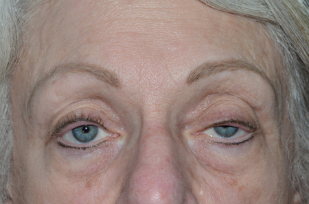
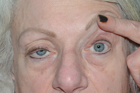
- In unilateral cases, check for presence of Hering’s phenomenon (Figures 1 and 2)

Figure 1. Bilateral ptosis worse on left with marginal reflex distance 1 mm on the right. Image courtesy Scott M. Goldstein, MD.

Figure 2. Ptosis on the right is worse with manual lid elevation on the left due to Hering’s law. Image courtesy Scott M. Goldstein, MD.
- Assess marginal reflex distance, lagophthalmos, levator function, crease height
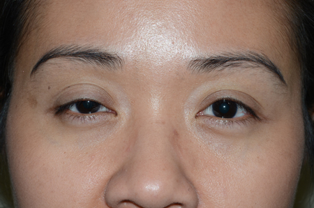
- Photographs for documentation (Figure 3)
- Consider provocative testing with phenylephrine to assess for Muller muscle function, where appropriate
- The four activities identified in Quality of Life assessment that improve after unilateral and bilateral surgery are the ability to perform fine manual work, reaching for objects above eye level, watching television and reading (Battu, 1996)

Figure 3. Ptosis. Image courtesy Scott M. Goldstein, MD.
- Visual field testing
- Quantifies degree of visual field loss and reversibility
- For some local carriers, the Center for Medicaid and Medicare Services (CMS) has defined a 30% loss of superior visual field as the minimum requirement for medically necessary ptosis repair
- Typically, height of superior visual field at 90-degree meridian is used to define superior visual field
- Decrease in field correlates with degree of ptosis (Meyer, Archives of Ophthalmology, 1989)
- Both manual kinetic and automated static visual field tests reliably document degree of impairment, but manual kinetic testing is faster and more sensitive (Rienmann, Archives of Ophthalmology, 2000)
- Assess ocular surface by slit-lamp biomicroscopy
- Schirmer testing
- Aponeurotic ptosis can be repaired in the context of dry eye condition that is being effectively controlled
- Widening the fissure can increase evaporation of the tear film and can worsen dry-eye syndrome (Mehta, American Journal of Ophthalmology, 2008)
- Tear volume may decrease after ptosis repair (Watanabe, Cornea, 2014)
Differential diagnosis
- Acquired myogenic ptosis
- Horner syndrome
- Previously unrecognized congenital ptosis
- Pseudoptosis
- Brow ptosis
- Dermatochalasis
- Hypertropia or contralateral hypoglobus
- Contralateral eyelid retraction
Management
- Levator aponeurosis advancement
- Anterior approach
- Local anesthesia is preferred, minimal sedation facilitates intra-operative monitoring
- Original description did not include dissecting aponeurosis off tarsus – in essence it was imbricated
- Suture material can be permanent or absorbable, braided or monofilament – original description by Jones was with 5-0 Chromic suture
- Outcomes
- 65%–77% success on first attempt (McCulley, OPRS, 2003; Scoppettuolo, British Journal of Ophthalmology, 2008)
- Bilateral and severe ptosis are indicators of higher reoperation rates
- Small-incision anterior approach
- Less-invasive approach (Lucarelli, American Journal of Ophthalmology, 1999)
- There have been modifications but as originally described an 8 mm incision is made at the lid crease facilitating a 10 mm dissection along the superior half of the tarsus
- The anterior surface of the aponeurosis is explored fully to a point several millimeters above the muscle aponeurotic junction
- A single central suture, or a second suture in close proximity is considered adequate support for this aponeurotic advancement
- The presumption of this technique is that more rapid and reliable healing can be expected with less tissue manipulation
- Dissection of the levator beyond the center 10 mm does not improve the post-operative result
- Release of the aponeurosis along the full width of its attachment to the tarsus is not needed
- In a study of 83 eyelids, when patients had good levator function surgical success was similar between small incision (n=18 eyelids, 94.7%) vs. standard incision (n= 20 eyelids, 95.2%). In patients with moderate levator function, surgical success was achieved less often with a small incision (n=14 eyelids, 66.7%) vs. standard incision (n= 18 eyelids, 81.8%) (Ranno, 2014)
- Unilateral ptosis repair by anterior levator advancement
- 27%–72% rate of postoperative symmetry (Tucker, Ophthalmology, 1999; Bartley, Transactions of the American Ophthalmological Society, 1996; Sarver, Archives of Ophthalmology, 1985)
- Assess for preoperative Hering phenomenon
- Some patients exhibit a contralateral intraoperative descent of the eyelid that resolves postoperatively
- Aim to position the operated eyelid to match the preoperative contralateral marginal reflex distance (Wladis, OPRS, 2008)
- Posterior approach
- Less common procedure
- Advances levator aponeurosis (“white line”) through a transconjunctival dissection (Patel, British Journal of Ophthalmology, 2010)
- Similar success rates to those published for anterior approach (Malhotra, Orbit, 2012)
- Muller muscle conjunctival resection
- Developed by Putterman and Urist in 1975 (Putterman, Archives of Ophthalmology, 1975)
- Several studies suggest that mullerectomy is a form of levator advancement (Marcet, OPRS, 2010; Glatt, Ophthalmic Surgery, 1990; Dresner, OPRS, 1991)
- Can be performed under general or local anesthesia because intra-operative monitoring is less critical
- Frontal nerve block can be used to avoid tissue swelling
- Epinephrine is omitted to prevent contraction of muller’s muscle
- Provocative testing can be performed pre-operatively with phenylephrine 2.5% drops
- Drops reflect the effect of resecting 8.25 mm of tissue
- Per classic description, adjust by 1 mm of tissue for each desired 0.5-mm change in eyelid position desired (Shields, Current Opinion in Otolaryngology & Head and Neck Surgery, 2003)
- Desmarres retractor is used to evert the lid
- Proposed advantages over levator advancement (Allen, Current Opinion in Ophthalmology, 2011)
- Avoids loss of eyelid sensation
- Lower reoperation rate in selected cases (< 2 mm of ptosis with good response to phenylephrine)
- Rapid recovery
- No requirement for intraoperative cooperation
- Proposed disadvantages versus levator advancement
- Less elevation
- Dermatochalasis cannot be addressed from a posterior approach.
- Theoretical consideration of damage to tear production
- Levator advancement versus mullerectomy
- One retrospective comparison
- High success rate in both groups
- Greater preoperative ptosis in levator advancement group
- Lower reoperation and better cosmetic outcomes with mullerectomy (Ben Simon, American Journal of Ophthalmology, 2005)
- Recent survey indicated that the majority of surgeons performed posterior ptosis repair, although levator advancement was the preferred technique when ptosis repair was performed in conjunction with blepharoplasty (Aakalu, OPRS, 2011)
- Hybrid Technique
- As an alternative to either the anterior levator aponeurotic advancement or posterior Mullerectomy, a hybrid technique can be performed (presented by Simeon Lauer at the ASOPRS Spring meeting, Newport, Rhode Island, 2013)
- The surgery is performed through a skin incision
- Levator aponeurosis is dissected from the anterior tarsal surface
- Sutures are passed to tighten Muller’s muscle and its the levator attachment to the superior tarsal border and the levator attachment to the anterior tarsal surface, to varying degrees depending on surgeon preference
- This technique offers more flexibility, requires experience as a ptosis surgeon
- There is a learning curve as the surgeon learns how tissues respond, intra-operative monitoring provides a good sense of how to balance the two areas of tightening the levator
Preventing and managing treatment complications
- Overview
- Complications of aponeurotic ptosis surgery are divided between the more serious issues that threaten ocular function versus outcomes which do not threaten ocular integrity but fail to meet expectations
- Unexpected outcomes can be further subdivided between those that are readily addressed by secondary surgery and those that are more difficult to correct
- Timing is an issue because some outcomes are best reversed immediately, others require a period of observation to be sure that it will not correct spontaneously while others are best corrected after allowing time for tissue healing
- Location is another issue because some unexpected outcomes are best addressed in the office or in a minor surgery room while other problems require a return trip to the operating room
- All of these issues play into the dynamic of a potentially compromised doctor patient relationship and possible medicolegal liability
- Ptosis in the context of trabeculectomy is difficult to correct by aponeurotic surgery. Bleb revision may be needed to improve the vector of the lid relative to the bleb (Clark, 2016)
- Overcorrection
- Spans a spectrum from subtle asymmetry which may not meet expectations to more significant overcorrection with incomplete lid closure, exposure keratopathy and nocuturnal lagophthalmos
- When the lid has been positioned at its desired height during surgery, usually with intra-operative monitoring, overcorrection is unexpected because gravity should pull the lid further down during healing
- The lid may have appeared lower during surgery than its actual position due to injected local anesthesia, sedation, blood and positioning
- Adhesions can develop between the site of aponeurotic repair and more superior structures such as the lacrimal gland, trochlea, or septum which can elevate the lid margin beyond the anticipated position
- Massage is usually the first proposed treatment because the problem may correct itself due to the pull of gravity
- Lubrication of the cornea is an effective temporizing measure
- The need for lubrication can also become a focus of concern as patients wonder about the timing of and need for secondary surgery
- Consider early revision if it is likely that an anatomic revision is needed
- Temporizing measures are better options when the lid is anatomically in good position (i.e. the problem is primarily due to the healing process) because early secondary surgery may exacerbate the aberrant healing
- Undercorrection
- When the surgeon is confident that the levator has been correctly positioned it is best to await resolution of post-operative edema rather than re-explore the lid
- Revision surgery may be necessary when the lid height is not responding to the aponeurotic repair as expected and the surgeon deems it necessary to create a different degree of muscle tightening
- Higher revision rates may be seen in patients with floppy eyelid syndrome (Nirmalan, OPRS 2024)
- Asymmetry
- Variable combination of over and under correction
- Parameters for surgical correction are as described above
- Ocular surface disease
- Lubricate with rewetting drops
- Consider ointment at bedtime
- Flip the eyelid over and assure that there is no exposed suture
- Infection
- 04% for eyelid outpatient procedures (Lee, OPRS, 2009)
- Manage with topical and oral antibiotics
- Induced astigmatism
- Mean change of 0.25 diopters (Zinkernagel, Archives of Ophthalmology, 2007)
- 8% developed increased with-the-rule astigmatism; 13.8% developed increased against-the rule astigmatism by 6 week postoperative interval (Holck, OPRS, 1998)
- By 12-month postoperative period, regressed in 100% of patients (Holck, OPRS, 1998)
- History
- Anterior apoenurotic repair was presented at the American Ptosis Society meeting in Dallas in September, 1973 by Dr. Marvin Quickert
References and additional resources
- Aakalu VK, Setabutr P. Current ptosis management: a national survey of ASOPRS members. Ophthal Plast Reconstr Surg. 2011 Jul-Aug;27(4):270-6.
- Ahmadi AJ, Saari JC, Mozaffarian D, et al. Decreased Carotenoid Content in Preaponeurotic Orbital Fat of Patients With Involutional Ptosis. Ophthal Plast Reconstr Surg, 21: 46-51, 2005.
- Allen RC, Saylor MA, Nerad JA. The current state of ptosis repair: a comparison of internal and external approaches. Curr Opin Ophthalmol, 22: 394-9, 2011.
- Altieri M, Truscott E, Kingston AE, et al. Ptosis secondary to anterior segment surgery and its repair in a two-year follow-up study. Ophthalmologica 2005; 219:129-135.
- Bartley GB, Lowry JC, Hodge DO. Results of levator-advancement blepharoptosis repair using a standard protocol: effect of epinephrine-induced eyelid position change. Trans Am Ophthalmol Soc 1996;94:165–73
- Battu VK1, Meyer DR, Wobig JL. Improvement in subjective visual function and quality of life outcome measures after blepharoptosis surgery. Am J Ophthalmol. 1996 Jun;121(6):677-86.
- Ben Simon GJ, Lee S, Schwarcz RM, et al. External levator advancement vs. Müller’s muscle-conjunctival resection for correction of upper eyelid involutional ptosis. Am J Ophthalmol 2005; 140:426–432.
- Bleyen I1, Hiemstra CA, Devogelaere T, van den Bosch WA, Wubbels RJ, Paridaens DA. Not only hard contact lens wear but also soft contact lens wear may be associated with blepharoptosis. Can J Ophthalmol. 2011 Aug;46(4):333-6.
- Cahill KV1, Bradley EA, Meyer DR, Custer PL, Holck DE, Marcet MM, Mawn LA. Functional indications for upper eyelid ptosis and blepharoplasty surgery: a report by the American Academy of Ophthalmology. Ophthalmology. 2011 Dec;118(12):2510-7.
- Cetinkaya A1, Kersten RC. Surgical outcomes in patients with bilateral ptosis and Hering’s dependence. Ophthalmology. 2012 Feb;119(2):376-81.
- Clark TJE, Rao K, Quinn CD, et al. A force vector model of upper eyelid position in the setting of a trabeculectomy bleb. Ophthal Plast Reconstr Surg 2016; 32, 127-132.
- Dortzbach RK, Sutula FC. Involutional blepharoptosis: a histopathological study. Arch Ophthalmol 1980;98:2045–9.
- Dresner SC. Further modifications of Müller’s muscle conjunctival resection procedure for blepharoptosis. Ophthal Plast Reconstr Surg 1991; 7:114–122.
- Fujiwara T1, Matsuo K, Kondoh S, Yuzuriha S. Etiology and pathogenesis of aponeurotic blepharoptosis. Ann Plast Surg. 2001 Jan;46(1):29-35.
- Glatt HJ, Putterman AM, Fett DR. Muller’s muscle-conjunctival resection procedure in the treatment of ptosis in Horner’s syndrome. Ophthalmic Surg 1990; 21:93–96.
- Holck DE1, Dutton JJ, Wehrly SR. Changes in astigmatism after ptosis surgery measured by corneal topography. Ophthal Plast Reconstr Surg. 1998 May;14(3):151-8.
- Jones LR, Quickert MH, Wobig JL. The cure of ptosis by aponeurotic repair. Arch Ophthalmol 1975; 93:629–634.
- Kakizaki H, Zako M, Ide A, et al. Causes of undercorrection of medial palpebral fissures in blepharoptosis surgery. Ophthal Plast Reconstr Surg 2004; 20:198-201.
- Kase S1, Noda M, Yoshikawa H, Yamamoto T, Ishijima K, Ishida S. Oxidative Stress in the Levator Aponeurosis in Asian Involutional Blepharoptosis. Ophthal Plast Reconstr Surg. 2014 May 14.
- Kersten RC1, de Conciliis C, Kulwin DR. Acquired ptosis in the young and middle-aged adult population. Ophthalmology. 1995 Jun;102(6):924-8.
- Kitazawa T. Hard contact lens wear and the risk of acquired blepharoptosis: a case-control study. Eplasty. 2013 Jun 19;13:e30.
- Lam BL, Lam S, Walls RC. Prevalence of palpebral asymmetry in white persons. Am J Ophthalmol 1995; 120:518-522
- Lee EW1, Holtebeck AC, Harrison AR. Infection rates in outpatient eyelid surgery. Ophthal Plast Reconstr Surg. 2009 Mar-Apr;25(2):109-10.
- Linberg JV, McDonald MB, Safir A, Googe J. Ptosis following radial keratotomy. Performed using a rigid eyelid speculum. Ophthalmology 1986; 93:1509
- Lucarelli MJ, Lemke BN. Small incision external levator repair: technique and early results. Am J Ophthalmol 1999; 127:637-644.
- Malhotra R, Salam A. Outcomes of Adult Aponeurotic Ptosis Repair Under General Anaesthesia by a Posterior Approach White-Line Levator Advancement. Orbit. 2012 Feb;31(1):7-12.
- Marcet MM1, Meyer DR, Greenwald MJ, Roth S, Selva D. Proximal tarsal attachments of the levator aponeurosis: implications for blepharoptosis repair. Ophthalmology. 2013 Sep;120(9):1924-9.
- Marcet MM, Setabutr P, Lemke BN, et al. Surgical microanatomy of the Müller muscle-conjunctival resection ptosis procedure. Ophthal Plast Reconstr Surg 2010; 26:360–364.
- McCulley TJ1, Kersten RC, Kulwin DR, Feuer WJ. Outcome and influencing factors of external levator palpebrae superioris aponeurosis advancement for blepharoptosis. Ophthal Plast Reconstr Surg. 2003 Sep;19(5):388-93.
- Medicare Local Coverage Determination for Blepharoplasty, Blepharoptosis, and Brow Lift (Effective 8/18/2008–5/1/2013). http://www.cms.gov/medicare-coverage-database/details/lcd-details.aspx?LCDId=27474&ContrId=161&ver=59&ContrVer=2&Date=08%2f26%2f2013&DocID=L27474&bc=AAAAAAgAIAAAAA%3d%3d&.
- Mehat MS, Sood V, and Madge S. Blepharoptosis Following Anterior Segment Surgery: a New Theory for an Old Problem. Orbit, 31: 274-8, 2012.
- Mehta VJ, Perry JD. Blepharoptosis repair outcomes from trainee versus experienced staff as the primary surgeon. Am J Ophthalmol. 2013 Feb;155(2):397-403
- Meyer DR, Linberg JV, Powell SR, et al. Quantitating the superior visual field loss associated with ptosis. Arch Ophthalmol. 1989;107:840–3.
- Nirmalan A, Tran MT, Tailor P, Hodge D, Bradley EA, Wagner LH, Bartley GB, Tooley AA. Obstructive Sleep Apnea Associated With Increased Failure Rate of Ptosis Repair. Ophthalmic Plast Reconstr Surg. 2024 Mar-Apr 01;40(2):201-205.
- Naseem M, Donker DL, Paridaens D. Blepharoptosis as a sign of severe Graves’ orbitopathy. Eye (Lond). 2009 Aug;23(8):1743-4.
- Patel V, Salam A, Malhotra R. Posterior approach white line advancement ptosis repair: the evolving posterior approach to ptosis surgery. Br J Ophthalmol 2010;94:1513–1518.
Pereira LS1, Hwang TN, Kersten RC, Ray K, McCulley TJ. Levator Superioris Muscle Function in Involutional Blepharoptosis. Am J Ophthalmol. 2008 Jun;145(6):1095-1098 - Putterman AM, Urist M: Müller’s muscle-conjunctival resection: technique for treatment of blepharoptosis. Arch Ophthalmol 1975, 93:619–623.
- Puvanachandra N1, Hustler A, Seah LL, Tyers AG. The incidence of ptosis following extracapsular and phacoemulsification surgery: comparison of two prospective studies and review of the literature. Orbit. 2010 Dec;29(6):321-3
- Ranno S, Sacchi M, Gonzalez MO, et al. Evaluation of levator function for efficacy of minimally invasive and standard techniques for Involutional ptosis. Am J Ophthalmol 2014; 157:209-213.
- Riemann CD, Hanson S, Foster JA. A comparison of manual kinetic and automated static perimetry in obtaining ptosis fields. Arch Ophthalmol. 2000;118:65–9
- Sarver BL, Putterman AM. Margin limbal distance to determine amount of levator resection. Arch Ophthalmol 1985;103: 354–6.
- Scoppettuolo E, Chadha V, Bunce C, et al. British Oculoplastic Surgery Society (BOPSS) National Ptosis Survey. Br J Ophthalmol. 2008 Aug;92(8):1134-8.
- Shields M and Putterman AC. Current Opinion in Otolaryngology & Head and Neck Surgery. 11: 261-6, 2003.
- Song MS1, Shin DH, Spoor TC. Incidence of ptosis following trabeculectomy: a comparative study. Korean J Ophthalmol. 1996 Dec;10(2):97-103.
- Sridharan GV, Tallis RC, Leatherbarrow B, Forman WM. A community survey of ptosis of the eyelid and pupil size of elderly people. Age Ageing 1995;24:21–4.
- Tucker SM, Verhulst SJ. Stabilization of eyelid height after aponeurotic ptosis repair. Ophthalmology 1999;106:517–22.
- van den Bosch WA, Lemij HG. Blepharoptosis induced by prolonged hard contact lens wear. Ophthalmology 1992; 99:1759
- Watanabe A1, Kakizaki H, Selva D, Ohmae M, Yokoi N, Wakimasu K, Kimura N, Kinoshita S. Short-term changes in tear volume after blepharoptosis repair. Cornea. 2014 Jan;33(1):14-7.
- Wladis EJ and Gausas RE. Transient Descent of the Contralateral Eyelid in Unilateral Ptosis Surgery. 24: 348-51, 2008.
- Zinkernagel MS1, Ebneter A, Ammann-Rauch D. Effect of upper eyelid surgery on corneal topography. Arch Ophthalmol. 2007 Dec;125(12):1610-2.
