Congenital Entropion
Updated May 2024
Cat N. Burkat, MD, FACS; Ann Tran, MD
Establishing the diagnosis
Definition
Inward rotation of the lower or upper eyelid margin, present at or shortly after birth (Maman, Ann Plast Surg 2011)
Etiology
- Proposed mechanisms of congenital upper eyelid entropion
- Direct mechanical pressure in utero
- Orbicularis spasm and hypertrophy from corneal abrasion
- Intrauterine tarsal deformity
- Proposed mechanisms of congenital lower eyelid entropion
- Vertical deficiency in the posterior eyelid lamella with or without disinsertion of the lower eyelid retractor muscles
- Horizontal laxity of the medial or lateral canthal tendons
- Facial nerve palsies
Epidemiology
- Congenital lower eyelid entropion is more common than upper eyelid entropion; however, both presentations are extremely rare.
- In a case series by Alsuhaibani et al. (Eye 2012), children with isolated facial nerve palsy were more prone to develop lower eyelid entropion; in contrast, adults with facial nerve palsy typically develop ectropion.
- Sires et al. (Ophthal Plast Reconstr Surg 1999) reported a retrospective clinical series of 25 cases of congenital horizontal tarsal kink. The classic presentation was male, Caucasian, diagnosed at 7.2 weeks of age, bilateral, or right-sided. Corneal ulcers were seen in 50% of patients.
History
- Typically presents at birth with diagnosis based on clinical malposition of eyelids
- Infrequent opening of eyes
- Photosensitivity
- Irritability
- Blepharospasm
- Corneal epithelial defects
Clinical features
- Upper eyelid congenital entropion
- More severe form of congenital upper eyelid entropion, known as the tarsal kink syndrome, is due to a congenital malformation of the tarsal plate. On eversion, the posterior tarsus demonstrates a large horizontal kinking (scarring) of the tarsus, that results in inversion of the eyelid margin and lashes onto the ocular surface.
- Typically absent upper eyelid folds (Biglan, Am J Ophthalmol 1980).
- Has been associated with cutis laxa, a rare connective disorder with redundant skin (Al-Faky, Pediatr Dermatol 2014).
- Lower eyelid congenital entropion
- Much more common than upper eyelid congenital entropion.
- Easier to diagnosis as more obvious clinical presentation at birth
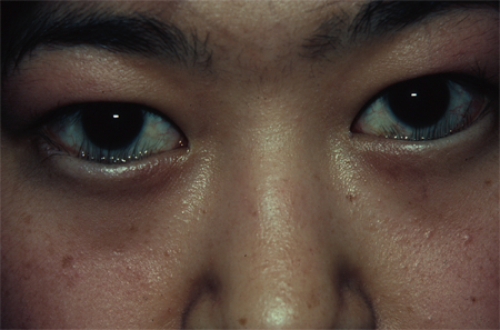
- Often confused with epiblepharon

Figure 1. Congenital entropion. Courtesy Rona Z. Silkiss, MD, FACS.
Risk factors
- A genetic predisposition has been suggested
- There are two case reports in the literature of neonates with bilateral congenital upper eyelid entropion and neonatal progeroid syndrome: aged face, wrinkled skin, decreased subcutaneous fat and neonatal teeth (Yazici, Ophthal Plast Reconstr Surg 2014). The entropion is believed to be secondary to abnormal activity of the orbicularis muscle, and a defective tarsal plate and retractor muscles.
Differential diagnosis
- Congenital epiblepharon: commonly seen in patients of Asian descent in which the eyelid margin is not truly inverted, but a hypertrophic fold of lower lid skin and orbicularis muscle override the lid margin and cause the lashes to rotate inwards. Pulling the fold of skin down gently can often demonstrate that the underlying eyelid margin is in proper anatomic position. Some patients with epiblepharon can outgrow the lid malposition as the midfacial bones and soft tissues mature.
- Distichiasis
- Trichiasis
- Trachoma
- Cicatrizing disorders
- Other types of entropion: cicatricial, involutional, acute spastic
- Can be confused with eyelid retraction
Patient management: treatment and follow-up
Natural history
- In a large case series of congenital horizontal tarsal kink, amblyopia developed in 4/15 patients, not related to corneal opacification (Sires, Ophthal Plast Reconstr Surg 1999).
- Poor visual prognosis is more likely to occur from a corneal ulcer scar or delay in diagnosis.
Medical therapy
- Symptomatic management and prevention of corneal irritation can reduce the progression of keratopathy
- Ocular lubricating ointment
- Frequent artificial tears
- Topical antibiotics if indicated
- Placement of steri-strips or tape to temporarily evert the eyelid margin
- Botulinum toxin: 5 units of botulinum toxin has been injected in the pretarsal orbicularis with no recurrence for up to 7 months in a case report of a 3-week old infant (Christiansen, Am J Ophthalmol 2004).
- Definitive surgery is often necessary to correct congenital entropion
Surgery
- Congenital upper eyelid entropion — multiple options:
- Lateral tightening if horizontal eyelid laxity is present
- Celsus procedure involves clamping and excising an elliptical area of skin from the anterior lamella (skin)
- Commonly used in veterinary medicine
- Does not address tarsal fibrosis/abnormalities
- Full-thickness blepharotomy through the eyelid at about 3 mm above the lash line, with suturing of inferior skin edge to the upper portion of tarsus to rotate the margin
- Transverse blepharotomy and tarsotomy at the deformed tarsal area (Streatfeild 1861, Panas 1888) without closure of defect
- Tarsal kink wedge resection from middle of tarsus rather than near lash line
- Modification using suture to close the tarsal wedge area, with the sutures brought out through the skin just above the lash line and tied over glass beads (Snellen 1862)
- Further modified to use a continuous suture to close the tarsal wedge area (Kiep 1924)
- Lamellar tarsoplasty modification (MacCarthy, Ophthalmic Surg 1984)
- Additional excision of orbicularis muscle overlying tarsal abnormality (Busacca 1936)
- Tarsal resection and placement of scleral graft as spacer
- Levator aponeurosis reinsertion
- Dailey et al. (Ophthal Plast Reconstr Surg 1999) reported one case of congenital entropion that was caused by levator disinsertion. Surgery to repair the levator aponeurosis disinsertion corrected the entropion.
- Anterior lamellar repositioning
- Price and Collin (Br J Ophthalmol 1987) described a technique in which the tarsal kink was corrected in one patient by incising the eyelid crease, and undermining the skin and orbicularis muscle to the lash roots. 6-0 polyglycolic acid sutures were passed through the skin just above the lashes, and secured higher up on the tarsus to provide lid eversion. Additional disinsertion of levator and Muller’s muscles helped weaken eyelid retraction, followed by suturing to the anterior lamella of the skin crease incision to further rotate the eyelashes.
- Transconjunctival horizontal tarsotomy with marginal rotation
- Naik et al. (Ophthalmology 2007) reported in a small retrospective case series that 5/5 patients had permanent correction of entropion after an average 10 month follow-up.
- Congenital lower eyelid entropion
- Surgical procedures for lower eyelid congenital entropion are similar to those for lower eyelid involutional entropion.
- Temporizing methods with Quickert-Rathbun sutures
- Posterior advancement of lower eyelid retractors (Takahashi, Orbit 2014)
- Lateral tarsal strip procedure (horizontal tightening of eyelid)
- Rotational sutures versus incisional surgeries were seen to have similar outcomes in a small case series of ten patients by Serafino et al. (Eur J Ophthalmol 2005). Rotational sutures were preferred given the simplicity and quickness of the procedure.
Complications
- Infectious keratitis
- Corneal abrasion
- Corneal ulceration (Lippincott, Trans Am Ophthalmol Soc 1894)
- Stromal opacification
- Amblyopia with permanent vision loss
- Early recognition and treatment improves visual prognosis
References and additional resources
- Al-Faky YH, Salih MA, Mubarak M, Al-Rikabi AC. Bilateral congenital entropion with cutis laxa. Pediatr Dermatol. 2014; 31: e82-84.
- Alsuhaibani AH, Bosley TM, Goldberg RA, Al-Faky YH. Entropion in children with isolated peripheral facial nerve paresis. Eye (Lond) 2012; 26: 1095-1098.
- Busacca A. Method of correction of entropion in trachomatous patients: with particular regard to esthetic results. Tr Ophth Soc U Kingdom. 1890; 16: 822-828.
- Biglan AW, Buerger GF, Jr. Congenital horizontal tarsal kink. Am J Ophthalmol. 1980; 89: 522-524.
- Christiansen G, Mohney BG, Baratz KH, Bradley EA. Botulinum toxin for the treatment of congenital entropion. Am J Ophthalmol. 2004; 138: 153-155.
- Dailey RA, Harrison AR, Hildebrand PL, Wobig JL. Levator aponeurosis disinsertion in congenital entropion of the upper eyelid. Ophthal Plast Reconstr Surg. 1999; 15: 360-362.
- Kiep WH. Modification of Snellen’s operation for entropion and trichiasis. Tr Ophthal Soc U Kingdom. 1924; 44: 372-375.
- Lippincott JA. Case of entropion, probably congenital, complicated with extensive ulceration of both corneae. Trans Am Ophthalmol Soc. 1894; 7: 225-226.
- Maman DY, Taub PJ. Congenital entropion. Ann Plast Surg. 2011; 66: 351-353.
- McCarthy RW. Lamellar tarsoplasty–a new technique for correction of horizontal tarsal kink. Ophthalmic Surg. 1984; 15: 859-860.
- Naik MN, Honavar SG, Bhaduri A, Linberg JV. Congenital horizontal tarsal kink: a single-center experience with 6 cases. Ophthalmology. 2007; 114: 1564-1568.
- Traitement operatoire de l’entropion granuleux. Bull Med Paris. 1888; 2: 827.
- Price NC, Collin JR. Congenital horizontal tarsal kink: a simple surgical correction. Br J Ophthalmol. 1987; 71: 204-206.
- Serafino M, Bottoli A, Nucci P. Correction of congenital entropion of the lower eyelid: incisional versus rotational surgery. Eur J Ophthalmol. 2005; 15: 536-540.
- Sires BS. Congenital horizontal tarsal kink: clinical characteristics from a large series. Ophthal Plast Reconstr Surg. 1999; 15: 355-359.
- Snellen H. Modification a l’operation de l’entropion. Cong Period Internat D’Ophthal. 1962; 2: 236-238.
- Streatfield JF. On grooving the fibro-cartilage of the lid in cases of entropion and trichiasis. Ophth Hosp Reports. 1858; 1: 121-130.
- Takahashi Y, Ikeda H, Ichinose A, Kakizaki H. Congenital entropion: outcome of posterior layer advancement of lower eyelid retractors and histological study of orbicularis oculi muscle hypertrophy. Orbit. 2014; 33: 444-448.
- Yazici B, Toka F, Comez AT. Anatomical characteristics and surgical treatment of bilateral congenital upper eyelid entropion in an infant with neonatal progeroid syndrome. Ophthal Plast Reconstr Surg. 2014; 30: e164-166.
