Fibrous Histiocytoma
Establishing the diagnosis
Etiology
- Arises from mesenchymal cells in fascia, muscle, or other soft tissues
- Orbit is the most commonly affected periocular structure
- Conjunctiva and eyelids can also be involved.
Epidemiology
- Three main subtypes. based on clinical behavior
- Benign 63%
- Locally aggressive 26%
- Malignant 11%
- A rare tumor, however the most common mesenchymal tumor of the orbit
- Most common in adults 30–60 years old
- 3%–10% occur in pediatric population; can occur following external beam radiation for retinoblastoma (Shields, Ophthal Plast Reconstr Surg. 2001)
- Male = female
- Some authors have suggested that fibrous histiocytoma refers specifically to fibroblastic spindle cell tumors of the orbit with aggressive behavior and have advocated re-classifying more superficial, benign lesions as dermatofibromas (Tieger, Ophthal Plast Reconstr Surg. 2019).
History
- Decreased vision
- Progressive proptosis
- Diplopia
- Previous history of FH
Clinical features
- Proptosis
- Dystopia
- Lid swelling
- Loss of vision
- Afferent pupillary defect due to compressive optic neuropathy
- Decreased motility
- Ptosis
- Chemosis
- Ephiphora and dacryocystitis if there is extension into the lacrimal sac (Stefanyszyn, Ophthal Plast Reconstr Surg. 1994)
- Most commonly occur supranasally, however can occur anywhere in the orbit and periorbital sctructures
- FH reportedly can occur in the lacrimal gland (Bajaj, Ophthal Plast Reconstr Surg. 2007)
Testing
Evaluation of motility, vision, pupils, Hertel exophthalmometry
Computed tomography
- Typically well circumscribed, but can be irregular
- Similar in appearance to schwannomas or cavernous hemangiomas
- Bone erosion is rare, but can be seen in recurrences or malignant lesions.
Magnetic resonance imaging
- T1
- Heterogenous
- Isointesnse to slightly hyperintense to muscle, hypointense to fat
- T2: Collagenous regions appear hypointense, whereas cellular areas are hyperintense.
- Heterogenous enhancement with gadolinium
Positron emission tomography
- Useful in detecting metastases as well as deciphering a recurrence from post-surgical changes (Char, Orbit. 2000)
Histopathology
- Benign:
- Poorly defined margins
- Spindle-shaped fibroblasts with a storiform pattern
- Can have vascular areas that are indistinguishable from hemangiopericytomas
- Locally aggressive
- Infiltrative margins
- Hypercellular
- Mitotic figures
- Malignant
- Infiltrative margins
- Nuclear pleomorphisms
- Atypia
- Necrosis
- Multinucleated giant cells
- Immunohistochemical staining
- Vimentin
- Alpha-antitrypsin
- Factor XIIIA
- Smooth muscle actin
- CD68

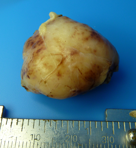
Figure 1. Completely excised fibrous histiocytoma.
Risk factors
- Middle aged adults
Differential diagnosis
- Cavernous hemangioma
- Hemangiopericytoma
- Schwannoma
- Solitary Fibrous tumor
Patient management: treatment and follow-up
Natural history
(Font, Hum Pathol. 1982.)
- 63% are benign, 26% are locally aggressive, and 11% are malignant.
- Recurrence:
- Benign: 31%
- Locally aggressive: 57%
- Malignant: 64%
- 10-year survival rates:
- Benign: 100%
- Locally aggressive: 92%
- Malignant: 23%
Medical therapy
- Radiation has been shown to be ineffective, and has even been reported to induce formation of malignant FH (Zhang, World J Surg Oncol. 2014; Shields, Ophthal Plast Reconstr Surg. 2001)
- Adjunctive chemotherapy
Surgery
- Local Excision for well-circumscribed lesions
- Locally invasive and malignant lesions often require a wide surgical excision.
- Malignant recurrences and aggressive lesions might require exenteration.
Preventing and managing treatment complications
- Surgical complications depend on technique and are discussed in greater detail in reviews of surgery.
- Chemotherapy complications should be managed by an oncologist.
Disease-related complications
- Recurrence
- Local invasion
- Intracranial invasion (Ueda, Neurol Med Chir (Tokyo). 2003)
- Metastasis
Patient instructions
Follow closely for recurrent orbital signs including decreased vision, pain, proptosis, diplopia or a palpable mass.
References and additional resources
- Bajaj MS, Pushker N, Kashyap S, Sen S, Vengayil S, Chaturvedi A. Fibrous histiocytoma of the lacrimal gland. Ophthal Plast Reconstr Surg. 2007 Mar-Apr;23(2):145-7.
- Black EH, et al. Smith and Nesi’s Ophthalmic Plastic and Reconstructive Surgery. 3rd ed. New York: Springer; 2012; 848-9.
- Cole SH, Ferry AP.Fibrous histiocytoma (fibrous xanthoma) of the lacrimal sac. Arch Ophthalmol. 1978 Sep;96(9):1647-9.
- Tieger MG, Jakobiec FA, Ma L, and Wolkow N. Small benign storiform fibrous tumor (fibrous histiocytomas) of the conjunctival substantia propria in a child: review and clarification of biologic behavior. Ophthalmic Plast Reconstr Surg. 2019; epub ahead of print.
- Stefanyszyn MA, Hidayat AA, Pe’er JJ, Flanagan JC. Lacrimal sac tumors. Ophthal Plast Reconstr Surg. 1994 Sep;10(3):169-84.
- Shields JA, Husson M, Shields CL, Krema H, Eagle RC Jr, Singh AD. Orbital malignant fibrous histiocytoma following irradiation for retinoblastoma. Ophthal Plast Reconstr Surg. 2001 Jan;17(1):58-61.
- Font RL, Hidayat AA. Fibrous histiocytoma of the orbit. A clinicopathologic study of 150 cases. Hum Pathol. 1982 Mar;13(3):199-209.
- Char D, Caputo G, Miller T. Orbital fibrous histiocytomas. Orbit. 2000 Sep;19(3):155-159.
- Ueda R1, Hayashi T, Kameyama K, Yoshida K, Kawase T. Orbital malignant fibrous histiocytoma with extension to the base of the skull–case report. Neurol Med Chir (Tokyo). 2003 May;43(5):263-6.
- Zhang GB, Li J, Zhang PF, Han LJ, Zhang JT. Radiation-induced malignant fibrous histiocytoma of the occipital: a case report. World J Surg Oncol. 2014 Apr 17;12:98
- AAO, Basic and Clinical Science Course. 2010-11.
- Garrity JA, Henderson JW. Henderson’s Orbital Tumors. 4th edition. Lippincott Williams & Wilkins. 2007.
- Rootman J. Diseases of the Orbit: A multidisciplinary approach. 2nd revised edition, Lippincott Williams & Wilkins. 2002.
