Canalicular Infections and Inflammation
Updated July 2024
Establishing the diagnosis
Etiology
Primary canaliculitis
- Infection secondary to bacteria, fungus, or virus
- Staphylococcus, Streptococcus, and Actinomyces were the most common microbes isolated (Pal, Seminal Ophthalmology, 2024).
- Primary canaliculitis occurs more frequently compared to secondary cannliculitis
- The inferior canaliculus is involved more commonly than superior.
- A number of other bacteria have also been implicated: Nocardia, Mycobacterium species, Staphylococcus, Streptococcus (Freedman, Surv Ophthalmol 2011; Zaldivar OPRS 2009).
- Bacteria are often among concretions, which are basophilic masses of amorphous material.
- Classic Actinomyces “sulfur” granules have an eosinophilic periphery with club-like structures (filamentous bacteria) (Freedman, Surv Ophthalmol 2011).
- Actinomyces is a facultative anaerobe; anaerobic cultures are required for isolation.
- Rarely, patients are found to have a canalicular diverticulum that can cause stasis and the propagation of bacteria (Adjemian, OPRS 2000).
- Staphylococcus is the most common organism cultured from specimens of canaliculitis (Anand, Orbit 2004; Vecsei, Ophthalmologica 1994).
Secondary canaliculitis
- Secondary infection after punctal or intracanalicular plug placement
- 8% incidence of canaliculitis with intra-canalicular plugs (Marcet, Ophthalmology 2015)
- Plugs can cause canalicular stasis
- Organisms isolated are often similar to primary canaliculitis (Freedman, Surv Ophthalmol 2011)
Epidemiology
- Mean age at presentation is approximately 60 years
- 5:1 female to male ratio
- In 1 study risk of canaliculitis after intracanalicular plug placement was about 5%
- Most often affects the lower eyelid (Vecsei, Ophthalmologica 1994)
History
Patients often have months to years of chronic symptoms, including discharge and irritation of the eye.
Determine whether patient has a history of punctal or intracanalicular plugs.
Clinical features
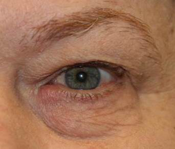
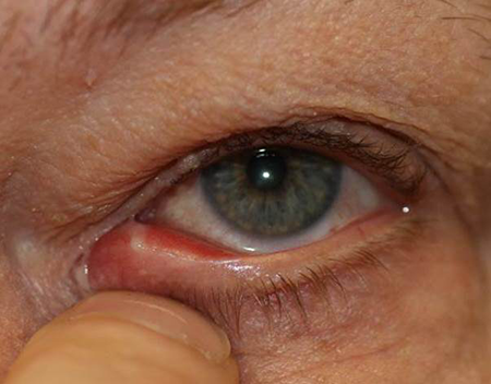
- Classic presenting signs (Figures 1–3)
- Epiphora
- Medial canthal edema
- Nonresolving conjunctivitis
- Occasionally patients are found to have a mass emanating from the punctum (papilloma or pyogenic granuloma) (Perumal, OPRS).

Figure 1. Canaliculitis stone. Courtesy Michael J. Hawes, MD.

Figure 2. Canaliculitis. Courtesy Gregory J. Griepentrog, MD.

Figure 3. Canaliculitis. Courtesy Gregory J. Griepentrog, MD.
Testing
- Slit-lamp examination: Edema of the medial eyelid, pouting punctum with discharge emanating from the punctum
- Attempt to express discharge from the punctum through compression over the medial eyelid
- Canalicular probe and irrigation
- Initial probe can be “gritty” secondary to canalicular concretions
- There may be some component of resistance to probing
- Irrigation often demonstrates a stenotic distal end of the canaliculus
- If classic signs of canaliculitis are present, probing and irrigation are likely not necessary
- Discharge can be sent for culture and/or histopathological analysis.
- Culture of punctal discharge
- Recommend culture be sent in separate aerobic and anaerobic transport media; Actinomyces is a facultative anaerobe
- Urgent transport of the material may increase yield
- Consider specifying culture for mycobacteria and fungi as well
- Histopathology of concretions or foreign body
- Consider hematoxylin and eosin (HE), Gram, Gomori’s methenamine silver (GMS), and Periodic acid-Schiff (PAS) stains to help identify the presence of granules and various bacteria and fungi (Repp, Ophthalmology 2009)
- Dacryocystography may be useful in atypical cases, potentially to diagnose a true canalicular diverticulum (Adjemian, OPRS 2000).
Risk factors
- Punctal or intracanalicular plug placement
- Abnormal anatomy (i.e., diverticulum) of the canalicular system
Differential diagnosis
- Chronic conjunctivitis
- Preseptal cellulitis
- Nasolacrimal duct obstruction with chronic dacryocystitis
- Hordeolum/chalazion of the medial eyelid
- Lacrimal sac tumor when in the lower eyelid
- Squamous cell carcinoma and mucoepidermoid carcinomas of the canaliculus (Charles, Arch Ophthalmol 2006)
Patient management: treatment and follow-up
Natural history
- Usually has a chronic course of mild to moderate conjunctivitis, irritation, and discharge
- Less likely to resolve with medical treatment alone; some type of surgical treatment is usually necessary (see below)
- Patients with a history of dry eye may not describe tearing
- Surgical management is needed in 74% of cases while medical management was done in 21% of cases (Pal, Seminal Ophthalmology, 2024)
Medical therapy
- Warm compresses, digital massage
- Topical antibiotics and steroids
- Oral antibiotics
- Expression of canalicular contents through the punctum with irrigation of antibiotics
- Penicillin
- Fortified cefazolin at a dose of 50 mg/ml was used in a recent study.
- Multiple irrigations may be necessary (Mohan, Indian J Ophthalmol 2008).
- Antibiotics may improve symptoms, though they are unlikely to lead to complete resolution; definitive treatment with medical therapy alone is more likely early in the course of disease.
- Hyperbaric oxygen has been used in refractory cases (Shauly, Graefes Arch Clin Exp Ophthalmol 1993).
- In secondary canaliculitis, it is recommended to remove the punctal plug if still in place, as the plug provides a mechanical obstruction.
Surgery
- The goal of surgical treatment is to remove the canalicular concretions through meticulous debridement along with any foreign body
- Dilation of the punctum with curettage of the canaliculus
- After dilation of the punctum, a curette can be placed in the canaliculus and contents can be expressed through the punctum (Freedman, Surv Ophthalmol 2011)
- Punctoplasty with curettage of the canaliculus
- Similar to above with use of a punctoplasty to improve access to the canaliculus
- Vertical canaliculotomy (modified punctoplasty) with retrograde expression of concretions
- Remove concretions through the punctum with the use of cotton-tipped applicators (Perumal, OPRS in press).
- Horizontal canaliculotomy through the conjunctiva with curettage; with or without closure of the canaliculus, with or without stent placement
- Probe is placed in the canaliculus and blade is used to make a horizontal incision through the conjunctiva or the skin along the probe
- Curette can be used to remove concretions
- The proximal 2–3 mm of canaliculus can be safely preserved
- Some surgeons advocate the placement of a stent after debridement of the canaliculus to prevent secondary closure (Khu, OPRS 2012)
- Reports of cannlicular obstruction post canaliculotomy without a stent have been reported to be as high as 20% (Wang, Current Eye Research 2021)
- Serial punctal dilation and non-incisional canalicular curettage can be used, however repeat curettage may be required (Bothra, Orbit 2020)
- Medical treatment with antibiotic and/or steroid drops is often continued after surgical treatment for 2–4 weeks
Common treatment responses, follow-up strategies
Patients can be followed on a weekly to monthly basis, depending on severity of symptoms.
Multiple surgical treatments may be necessary depending on the patient response.
- If this is the case, suspect a retained foreign body
Preventing and managing treatment complications
- Recurrence of infection due to inadequate treatment
- Canalicular scarring/stricture
- May be prevented by avoiding repeated probings and treatments if possible
- Recurrent irrigation may dislodge an existing plug and send it to a “downstream” location, thus necessitating more involved surgical interventions (including dacryocystorhinostomy)
Disease-related complications
- Canalicular stenosis
- Chronic epiphora
Historical perspective
Canaliculitis was discovered to be an infectious condition in 1854 by Von Graefe. Actinomyces species was found to be the most common cause at that time (Freedman, Surv Ophthalmol 2011)
References and additional resources
- Adjemian, A and Burnstine MA. Lacrimal canalicular diverticulum: a cause of epiphora and discharge. Ophthal Plast Reconstr Surg 2000;16:471-2.
- Anand S, Hollingworth K, Kumar V, Sandramouli S. Canaliculitis: the incidence of long-term epiphora following canaliculotomy. Orbit 2004;23:19-26.
- Bothra N, Sharma A, Bansal O, Ali MJ. Punctal dilatation and non-incisional canalicular curettage in the management of infectious canaliculitis. Orbit. 2020 Dec;39(6):408-412.
- Charles NC, Lisman RD, and Mittal KR. Carcinoma of the lacrimal canaliculus masquerading as canaliculitis. Arch Ophthlamol 2006;124: 414-6.
- Freedman JR, Markert MS, Cohen AJ. Primary and secondary lacrimal canaliculitis: a review of literature. Surv Ophthalmol 2011;56:336-47.
- Kaliki S, Ali MJ, Honavar SG, Chandrasekhar G, Naik MN. Primary canaliculitis: clinical features, microbiological profile, and management outcome. Ophthal Plast Reconstr Surg 2012;28:355-60.
- Khu J, Mancini R. Punctum-sparing canaliculotomy for the treatment of canaliculitis. Ophthal Plast Reconstr Surg 2012;28:63-65.
- Marcet MM, Shtein RM, Bradley EA, et al. Safety and efficacy of lacrimal drainage system plugs for dry eye syndrome: a report by the American Academy of Ophthalmology. Ophthalmology 2015;122:1681-7.
- Mohan ER, Kabra S, Udhay P, et al. Intracanalicular antibiotics may obviate the need for surgical management of chronic suppurative canaliculitis. Indian J Ophthalmol 2008;56:338–40.
- Pal SS, Alam MS. Lacrimal Canaliculitis: A Major Review. Semin Ophthalmol. 2024 May 19:1-9. doi: 10.1080/08820538.2024.2354689.
- Perumal B, Meyer DR. Vertical canaliculotomy with retrograde expression of concretions for the treatment of canaliculitis. Ophthalmic Plast Reconstr Surg. 2015 Mar-Apr;31(2):119-21.
- Repp DJ, Burkat CN, Lucarelli M. Lacrimal excretory system concretions: canalicular and lacrimal sac. Ophthalmology 2009;116:2230-5.
- Shauly Y, Nachum Z, Gdal-On M, et al. Adjunctive hyperbaric oxygen therapy for actinomycotic lacrimal canaliculitis. Graefes Arch Clin Exp Ophthalmol 1993;231:429-31.
- Vecsei VP, Huber-Spitzy V, Arocker-Mettinger E, Steinkogler FJ. Canaliculitis: difficulties in diagnosis, differential diagnosis and comparison between conservative and surgical treatment. Ophthalmologica 1994;208:314-7.
- Wang M, Cong R, Yu B. Outcomes of Canaliculotomy with and without Silicone Tube Intubation in Management of Primary Canaliculitis. Curr Eye Res. 2021 Dec;46(12):1812-1815.
- Zaldivar RA, Bradley EA. Primary canaliculitis. Ophthal Plast Reconstr Surg 2009;25:481-4.
