Lateral and Medial Canthoplasty
Updated July 2024
Goals
Restore normal anatomic eyelid position
- Correction of involutional lower lid ectropion, entropion, and/or retraction
Maintain anatomic apposition of lid to globe
- Lid laxity causes poor lid-to-globe apposition leading to tear film instability and exposure keratopathy
Restore normal lid tension
- Tear pump failure can result from excessive lid laxity or facial nerve palsy (Vick, OPRS 2004)
- Floppy eyelid syndrome, eyelid imbrication (see below)
Improve aesthetic appearance of eyelid
- Eyelid contour can be affected by laxity of lateral or medial canthal tendons
- Rounding of lateral commissure
- Contour abnormality can result from performing lower lid blepharoplasty without addressing lid laxity.
- Lateral canthal dystopia can be involutional, traumatic, or developmental
- Normal position of lateral commissure slightly higher than medial commissure
- Medial canthal deformities (e.g., telecanthus, dystopia, epicanthal folds) are usually traumatic or congenital
Indications and contraindications
Indications
Lid malposition due to horizontal laxity
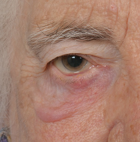
- Involutional ectropion (Bedran, Semin Ophthal 2010) (Figure 1)
- Poor lid-to-globe apposition causing exposure keratopathy
- Punctal ectropion causing epiphora
- Involutional entropion (Pereira, Semin Ophthal 2010)
- Significant ocular discomfort caused by lashes and keratinized skin rubbing directly on cornea
- Pathophysiology
- Lower-lid laxity
- Dehiscence of lower lid retractors
- Overriding orbicularis — often exacerbated by irritative symptoms causing blepharospasm (“spastic” entropion)
- Enophthalmos
- Lower-lid retraction (Chang OPRS 2011)
- Involutional — lid laxity
- Cicatricial — infection, inflammation, trauma, burns, postsurgical (e.g., lower-lid blepharoplasty, laser skin resurfacing)
- Mechanical — midface ptosis, craniofacial anomalies, tumor
- Paralytic — facial nerve palsy

Figure 1. Involutional right lower lid ectropion.
Tear pump failure
(Meyer, Curr Opin Ophthal 1993)
- Involutional and/or paralytic
Medial canthal tendon (MCT) laxity
- Severe laxity, especially in setting of facial nerve paralysis, can cause punctal ectropion, medial lower lid retraction, lagophthalmos/exposure keratopathy, and epiphora.
- Performing lateral canthal tendon (LCT) tightening in presence of MCT laxity can lateralize punctum and cause lacrimal outflow deficiency.
Canthal malposition
- Involutional, developmental, postsurgical, or traumatic
Floppy eyelid syndrome
(Culbertson, AJO 1981; Moscato, Compr Ophthal Update 2007)
- Marked lid laxity associated with softening of tarsus
- Multiple possible factors implicated in pathogenesis:
- Prone or side sleeping position causes mechanical pressure on lids
- Ischemia and reperfusion injury
- Upregulation of matrix metalloproteinases (MMP) implicated in elastin degeneration
- Lids can spontaneously evert during sleep, causing exposure keratopathy and chronic papillary conjunctivitis
- Associated with obstructive sleep apnea and obesity
- Surgical treatment involves upper-eyelid tightening
Eyelid imbrication
(Karesh, Ophthalmology 1993)
- Lid laxity causes upper-lid margin to overlap lower lid
- Upper palpebral conjunctiva rubs across lower lashes, leading to chronic irritation
- Sometimes associated with floppy eyelid syndrome
- Can be addressed with lower- and/or upper-lid tightening
Reconstruction following trauma or surgery
- Traumatic LCT/MCT avulsion
- Must rule out canalicular injury with MCT avulsion
- LCT resuspension following emergent lateral canthotomy and cantholysis for orbital compartment syndrome
- Tumor resection
Mild lower-lid laxity or lateral canthal deformity
- Open or closed lateral canthoplasty often performed in conjunction with various facial rejuvenation procedures (Taban, OPRS 2010) (e.g., upper- or lower-lid blepharoplasty, midface lift)
Contraindications
Relative
- Significant medial canthal tendon laxity (see above)
- Proptosis: surgical tightening of lids in presence of proptosis can lead to lid retraction
- Options for amelioration:
- Supraplacement of LCT on orbital rim (McCord 2008)
- Midface lift with possible malar augmentation to improve negative vector between cheek and orbit (Steinsapir, PRS 2003)
- Correction of proptosis (orbital decompression) or lateral rim repositioning (Kikkawa, Ophthalmology 2002)
Absolute
- Presence of malignancy, infection, or other pathology that would compromise surgical result
Preprocedure evaluation
Patient history
HPI
- Inquire about complaints of tearing, ocular discomfort, exposure symptoms.
- Tearing often multifactorial: excessive reflex tearing and deficient lacrimal outflow
Conditions that can cause or exacerbate involutional changes
- Facial nerve paralysis
- Bell’s palsy
- Facial trauma
- Surgery (e.g., vestibular schwannoma resection)
- CNS tumor
- Blepharospasm
- Obstructive sleep apnea (OSA)
- Risk factor for floppy eyelid syndrome (Moscato, Compr Ophthal Update 2007)
- Should be referred for sleep study if OSA suspected due to risk of systemic complications
Conditions that can cause or exacerbate cicatricial abnormalities
- Infection
- Trachoma
- HSV/HZV
- Necrotizing fasciitis
- Inflammation
- Stevens-Johnson syndrome
- Ocular cicatricial pemphigoid (OCP)
- Sarcoidosis
- Sezary syndrome, mycosis fungoides
- Tumors (primary or following resection/reconstruction)
- Basal cell carcinoma
- Squamous cell carcinoma
- Sebaceous carcinoma
- Melanoma
- Trauma
- Periorbital lacerations
- Burns (thermal or chemical)
- Surgery
- Lower-lid blepharoplasty
- Laser skin resurfacing
- Facial/orbital fracture repair
Conditions associated with medial or lateral canthal deformities
- Congenital anomalies (Jones 2013; Fries, Surv Ophthalm 1990)
- Down syndrome
- Craniosynostoses (Crouzon, Apert syndrome etc.)
- Euryblepharon
- Mandibulofacial dysostosis (Treacher-Collins syndrome)
- Oculoariculovertebral dysplasia (Goldenhar syndrome)
- Blepharophimosis syndrome
- Narrowed horizontal palpebral fissure
- Ptosis
- Epicanthus inversus
Trauma
- Naso-orbito-ethmoid (NOE) fractures (Markowitz, PRS 1991)
- Zygomaticomaxillary complex (ZMC) fractures
- Under-reduced frontozygomatic fracture (Converse, Clin Plast Surg 1975)
- Failure to resuspend midface following subperiosteal dissection, resulting in midface ptosis and lateral canthal dystopia (Lee 2008)
- Periocular lacerations
Clinical examination
Lid laxity
- Snap-back test, lid distraction
- MCT laxity: Distract lid laterally and observe punctal position
- Laxity significant if punctum moves close to or beyond nasal limbus
- Floppy eyelids
- Easily evertable upper lids
- Papillary conjunctivitis
- Eyelid imbrication: upper lid overlaps lower with lid closure
Lid malposition
- Ectropion, entropion, or retraction
- Punctal ectropion or override (“kissing puncta”)
Lagophthalmos
Retractor disinsertion
- Associated with involutional entropion and severe involutional ectropion
- With lower lid eversion, edge of disinserted retractor can be visualized subconjunctivally as a transition from white to pink below the inferior border of the tarsus.
Lamellar shortening
- Scarring or shortening of anterior or posterior lamella can cause ectropion or entropion, respectively.
- Scarring of the orbital septum (middle lamella) can cause tethering of the lid to the orbital rim.
Midface ptosis and/or hypoplasia
- Downward traction exerted by cheek on lower lid can exacerbate retraction and ectropion
- Malar hypoplasia (developmental or age-related) can create negative vector of forces apposing lid to globe.
Signs of corneal exposure
- Epithelial erosions/defects, scarring, etc.
Lacrimal outflow evaluation as indicated
- Primary dye test or dye disappearance test
- Canalicular probing and irrigation
Proptosis or enophthalmos
Preoperative assessment
Treatment must be individualized to each patient.
Involutional ectropion
- Distract lid laterally to estimate effect of tightening on lid position.
- Consider medial spindle in addition to lower lid tightening if punctal ectropion persists (Nowinski, Arch Ophthalmol 1985).
- Evaluate for possible retractor dehiscence.
Involutional entropion
- Repair typically involves lower lid tightening, retractor reinsertion, and excision of overriding orbicularis (if external approach used).
- Retractors can be approached transconjunctivally (Erb, Ophthalmology 2006) or externally through subciliary incision.
Lower-lid retraction
- Critical to distinguish involutional from cicatricial, mechanical, and paralytic causes
- Depending on underlying pathology, might need to consider adjunctive procedures in addition to lid repositioning
- Midface ptosis — midface lift
- Midface hypoplasia — malar augmentation
- Cicatricial entropion or thyroid orbitopathy — posterior lamellar spacer graft
- Anterior lamellar scarring — full thickness skin graft
- Scarring of orbital septum — dermis fat graft spacer Chang
Medial canthal tendon laxity
- MCT plication can be performed alone or concurrently with lateral canthoplasty
- Anterior crus can be plicated transcutaneously (Sodhi, J Cranio-Maxillofacial Surg 2005).
- Might not improve globe apposition
- Posterior plication can be performed through transcaruncular approach (Fante, OPRS 2001).
- Theoretical advantage in correcting globe apposition due to posterior vector
Lateral canthal dystopia
- Usually correctable with standard LCT suspension from lateral orbital wall, with or without lower lid tightening
- In recurrent or complex cases (e.g., cicatricial retraction, midface reconstruction), lateral canthopexy may be considered by securing with sutures or wires to:
- Drill holes in lateral rim
- Titanium screws
- Bone anchors (e.g., Mitek, Lactosorb syndrome, etc.)
Medial canthal dystopia
- Usually post-traumatic or developmental
- Can be addressed with medial canthopexy or Z-plasty reconstruction
Telecanthus
- Usually traumatic or congenital
- Normal intercanthal distance approximately equal to horizontal palpebral fissure length
- Must determine whether abnormality is purely soft tissue, or if underlying bony deformity is present (i.e., telorbitism or hypertelorism)
- Repair technique largely depends upon this anatomic distinction
Epicanthal folds
- Numerous options involving advancement or transposition flaps
- Must determine whether attachment of MCT is in proper anatomic position; medial canthopexy may be required if not
Procedure alternatives
Nonsurgical
Observation
Ocular lubrication
Corneal protection
- Burns (Malhotra Surv Ophth 2009), severe cicatrization
- Moisture chambers
- PROSE lens (Kalwerisky, Ophthalmology 2012)
- Amniotic membrane (ProKera) (Pachigolia, Eye Contact Lens 2009)
Horizontal lid taping
- Involutional entropion
Surgical
Lagophthalmos
- Tarsorrhaphy (McInnes, AJO 2006) (suture or permanent)
- Lid weight implantation (Mavrikakis OPRS 2006)
- Upper-lid recession (Ben Simon, AJO 2005)
- Full-thickness blepharotomy (Demirci OPRS 2007)
Involutional entropion
- Quickert sutures (Quickert, Arch Ophth 1971)
Lid tightening
- Full-thickness wedge resection (Callahan 1956) (largely historical)
Surgical techniques
Horizontal lower lid tightening
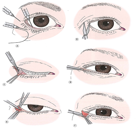
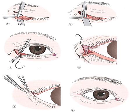
Lateral tarsal strip procedure
(Anderson Arch Ophth 1979) (Figure 2)
- Infiltrate local anesthetic.
- Perform lateral canthotomy and inferior cantholysis.
- Determine amount of horizontal laxity.
- Place lateral traction on lid and mark point where lid crosses lateral rim and commissure.
- Create tarsal strip
- Dissect anterior lamella and excise to point where lid crosses lateral commissure.
- Excise marginal epithelium.
- Detach retractors/conjunctiva from inferior edge.
- Remove palpebral conjunctiva with blade, low energy cautery, or radiofrequency ablation.
- Trim tarsal strip to point where lid crosses orbital rim
- Suspend strip from periosteum over inner aspect of rim
- 4-0 or 5-0 absorbable or nonabsorbable suture (e.g., polyglactin, polydiaxanone, or polypropylene) on a small half-circle needle (P-2 or OPS-5)
- Horizontal mattress or half-horizontal mattress pattern, ensuring positioning of strim posterior to lateral rim
- Slight overcorrection advisable: about 2–3 mm superior to intended final position of lateral commissure
- Reform lateral canthal angle (Weber, Ophth Surg 1991)
- Trim redundant skin
- Close skin


Figure 2. Lateral tarsal strip procedure. From Nerad JA, Techniques in Ophthalmic Plastic Surgery, 2010.
Modified Bick procedure
(Barrett, OPRS 2012)
- Lateral canthotomy/inferior cantholysis
- Distract lid laterally and mark point where lid crosses lateral rim.
- Excise triangular wedge of lateral canthal tendon and tarsus.
- Suspend end of tarsus to periosteum as above.
- Reform lateral canthal angle.
- Close skin.
Closed lateral canthoplasty
(Taban, Georgescu, Rizvi, Lessa)
- Carry dissection to lateral orbital rim through lateral upper lid crease incision.
- Pass suture internal to external through lateral commissure, then back internally at same point.
- Pass suture through periosteum behind lateral orbital rim and tie.
- Suture can also be passed through drill holes in lateral rim using “Leicester lasso” technique (Kannan OPRS 2014).
- 4-0 silk suture twisted into lasso and passed external to internal through drill holes x2
- LCT suture ends passed through lasso, pulled through drill holes with lasso, and tied
Reinforcement lateral canthoplasty
(Dailey OPRS 2011)
- For complex or recurrent LCT laxity/dehiscence
- Superior and inferior crus of LCT approached through supraciliary/subciliary incisions
- LCT plicated and suspended from periosteum behind lateral orbital rim
- Y-shaped graft (e.g., autogenous fascia lata, acellular dermal matix, porcine dermal collagen) sutured to limbs of LCT and periosteum over lateral rim
Adjunctive procedures
- Medial spindle procedure (Nowinski, Arch Ophthalmol 1985)
- Excision of diamond-shaped wedge of conjunctiva/retractors inferior to lower punctum
- Double-armed absorbable suture (e.g., 6-0 polyglactin or chromic gut) passed from inferior to superior wound edges, then through inferior fornix and externalized, tied over skin
- Orbitomalar ligament suspension (Korn PRS 2010)
- Dissection carried from lateral canthotomy through orbitomalar ligament under lateral midface, suture passed through deep aspect of SOOF and suspended from lateral orbital rim periosteum
- Alternative fixation technique: LCT suture passed through single drill hole in lateral rim and tied externally to SOOF suture (Oh, OPRS 2013)
- Midface lift
- Preperiosteal (Marshak OPRS 2010) or subperiosteal (Elner, Arch Fac Plast Surg 2003) dissection
- Consider malar augmentation for midface hypoplasia (Steinsapir, PRS 2003; Binder, Fac Plast Surg 2011)
- Midface tissues suspended and fixated with sutures to drill holes, screws, malar implants, or elevated with fixation devices (e.g., Endotine™ midface (Berkowitz, Aesth Surg J 2005)
Upper-lid tightening
- Full-thickness wedge resection (traditional)
- Lateral tarsal strip procedure or modified Bick procedure can be performed on upper lid in similar manner as lower lid (Dutton, AJO 1985; Perlman, OPRS 2002)
- 4-lid lateral tarsal strip-periosteal flap technique (Burkat, OPRS 2005)
- 5-mm lateral canthus incision to expose lateral rim
- 6-mm periosteal flap created and reflected medially
- Lateral tarsal strips fashioned in standard fashion and fixated to periosteal flaps with 5-0 polyglactin suture
Medial canthoplasty
Anterior medial canthal tendon plication
(Sodhi 2005)
- Create skin incisions (single horizontal or double vertical) over anterior crus of MCT and medial end of tarsus
- Pass suture sequentially through MCT near bony insertion, subcutaneous tunnel, and medial end of tarsus, taking care to avoid canaliculus (placement of lacrimal probe helpful)
- Tie under appropriate tension
- Close skin incisions(s)
Transcaruncular (posterior) medial canthal tendon plication
(Fante OPRS 2001)
- Incise conjunctiva below medial end of lower tarsus
- Create transcaruncular incision between caruncle and plica semilunaris, dissect bluntly to medial orbital wall
- Engage medial end of tarsus with suture (4-0 polypropylene on P-2 needle), avoiding canaliculus, and pass suture subconjunctivally to transcaruncular incision
- Pass same suture through periosteum at or above posterior lacrimal crest, then back through subconjunctival space to exit incision beneath tarsus
- If medial lower lid retraction is present, suture can be fixated more superiorly on medial rim Moe
- Adjust suture tension and tie
- Rotate knot to bury it in medial orbit
Medial tarsal suspension for medial lower lid retraction
(Frueh OPRS 2002)
- Create horizontal skin incisions from medial commissure to within 2 mm medial to each punctum.
- Dissect pockets in suborbicularis plane medial to lower punctum (avoiding canaliculi) and in superomedial upper lid.
- Pass suture (5-0 or 4-0 nonabsorbable) through periosteum over superomedial rim.
- Pass same suture through medial end of lower tarsus and tie under appropriate tension.
- Rotate knot to bury next to superomedial rim.
- Sew edges of upper and lower lid skin incisions together (medial tarsorrhaphy).
Repair of epicanthal folds
- Create canthoplasty incisions.
- Mustarde “stick man” (Anderson, Arch Ophth 1989)
- 5-flap technique
- Skin redraping method (Sa, Ophthalmology 2012)
- Undermine and transpose myocutaneous flaps.
- Excise excess soft tissue.
- Close skin and soft tissues.
Medial canthal z-plasty for MC dystopia
(Fox 1976)
- Create z-plasty incision incorporating anterior crus of MCT into lower limb of Z.
- Dissect MCT from lacrimal sac, undermine flaps.
- Transpose flaps and resuspend MCT in higher position with suture to periosteum over frontal process of maxilla.
- Close incisions.
Medial canthopexy
Congenital hypertelorism
- Bony reconstruction required
- Multidisciplinary craniofacial team approach recommended
Primary repair of NOE fracture
(See Orbital Fractures)
Transnasal wiring
(Smith 1992; Dutton, AJO 1985)
- Bilateral telecanthus, e.g., blepharophimosis (Sebastia, Aesth Plast Surg 2011) bilateral NOE fractures
- MCTs are wired to each other through drill holes across the nasal cavity.
- Wire can aid in reduction/fixation of bony abnormalities, or in setting of normal bony anatomy, can be passed through drill holes to engage MCTs and reduce telecanthus.
- Surgical approaches include bicoronal, Lynch incision, or incision directly over MCT (Nunery incision) (Timoney, OPRS 2012).
- Unilateral telecanthus (traumatic)
- MCT is wired across the nasal cavity to stable bone (frontal process of maxilla or frontal bone) on the opposite side (Kelly, OPRS 2004; Markowitz, PRS 1991).
- Titanium wire with barb and needle (Synthes®) can be used transnasally or anchored directly to frontal process (Engelstad Int J Oral Maxillofac Surg 2012).
Fixation of MCT to bone anchors
- Titanium microplates/screws (Shore, Ophthalmology 1992; Howard, Arch Ophth 1992)
- Mitek bone anchor (Antonyshyn, PRS 1996; Goldenberg, Ann Plast Surg 2008)
- Lactosorb anchor (Sharma, Arch Ophth 2006)
Patient management
- Postoperative instructions
- Appropriate activity limitations
- Keep wounds clean and dry.
- Monitor for signs of infection.
- Medications
- Antibiotic ointment to incisions
- Pain medications as needed
- Counsel patient that lids will be initially tight and will slowly relax over time.
- If nonabsorbable skin sutures used, remove 5–7 days postoperatively.
- Monitor for appropriate anatomic changes and functional/aesthetic response to treatment.
Complications
- Failure to correct underlying pathology
- Suture failure with canthal dehiscence
- Multiple sutures can be placed primarily to prevent
- Suture granulomas
- Treatment: excision, steroid injection
- Lateralization of punctum caused by unrecognized/unaddressed medial canthal tendon laxity
- Lid retraction can occur in setting of proptosis or aggressive lower lid blepharoplasty.
- Diplopia due to scarring of conjunctiva or lateral rectus tendon sheath
Disease-related complications
- Uncommon, usually associated with involutional changes
- Facial nerve palsy: exposure keratopathy, epiphora, brow ptosis
- Floppy eyelid syndrome: associated with obstructive sleep apnea
Historical perspective
Repair of involutional lower lid ectropion
- Traditional surgery involved full-thickness wedge resection.
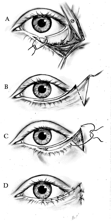
- Kuhnt-Szymanowski procedure (Callahan 1966) (Figure 3)
- Bick procedure (Bick, AJO 1963)
- Lateral tarsal strip procedure described by Anderson in 1979 (Anderson, Arch Ophth 1979)
- Many modifications of tarsal strip now used
Repair of involutional lower lid entropion
- Most early repair techniques focused on retractors and/or orbicularis.
- Jones, Reeh, and Tsukimura (Jones 2013)
- Quickert sutures
- Full-thickness wedge resection advocated by Hill, Quickert
- Lateral tarsal strip introduced and has become standard component of entropion repair

Figure 3. Kuhnt-Szymanowski procedure. From Callahan A. Reconstructive Surgery of the Eyelids and Ocular Adnexa, 1966.
References and additional resources
- Anderson RL, Gordy DD. The tarsal strip procedure. Arch Ophthalmol. 1979:97;2192.
- Anderson RL, Nowinski TS. The five-flap technique for blepharophimosis. Arch Ophthalmol. 1989;107:448-52.
- Antonyshyn OM, Weinberg MJ, Dagum AB. Use of a new anchoring device for tendon reinsertion in medial canthopexy. Plast Reconstr Surg 1996;98:520-23.
- Barrett RV, Meyer DR. The modified Bick quick strip for surgical treatment of eyelid malposition. Ophthal Plast Reconstr Surg 2012;28:294-9.
- Bedran EG, Periera MV, Bernardes TF. Ectropion. Semin Ophthalmol 2010;25:59-65.
- Ben Simon GJ, Mansury AM, Schwarcz RM, et al. Transconjunctival Müller muscle recession with levator disinsertion for correction of eyelid retraction associated with thyroid-related orbitopathy. Am J Ophthalmol 2005;140:94-99.
- Berkowitz RL, Apfelberg DB, Simeon S. Midface lift technique with use of a biodegradable device for tissue elevation and fixation. Aesthet Surg J 2005;25:376-82.
- Bick MW. Orbital tarsal disparity. Arch Ophthalmol 1966;75:386.
- Jones LT, Reeh MJ, Tsukimura JK. Senile entropion. Am J Ophthalmol. 1963;55:463-9.
- Binder WJ. Facial rejuvenation and volumization using implants. Facial Plast Surg 2011;27:86-97.
- Burkat CN, Lemke BN. Acquired lax eyelid syndrome: an unrecognized cause of chronic ocular irritation. Ophthal Plast Reconstr Surg 2005;21:52-8.
- Callahan A. Reconstructive Surgery of the Eyelids and Ocular Adnexa. Birmingham, Alabama: Aesculapius Publishing Co., 1966: 120-157.
- Chang EL, Rubin PA. Upper and lower eyelid retraction. Int Ophthalmol Clin 2002;42:45-59.
- Chang HS, Lee D, Taban M, Douglas RS, Goldberg RA. “En-glove” lysis of lower eyelid retractors with AlloDerm and dermis-fat grafts in lower eyelid retraction surgery. Ophthal Plast Reconstr Surg 2011;27:137-41.
- Converse JM, Smith B, Wood-Smith D. Deformities of the midface resulting from malunited orbital and naso-orbital fractures. Clin Plast Surg 1975;2:107-203.
- Culbertson WW, Ostler HB. The floppy eyelid syndrome. Am J Ophthalmol 1981;92:568-75.
- Dailey RA, Chavez MR. Lateral canthoplasty with acellular cadaveric dermal matrix graft (AlloDerm) reinforcement. Ophthal Plast Reconstr Surg 2011:28;e29-31.
- Danks JJ, Rose GE. Involutional lower lid entropion: to shorten or not to shorten? Ophthalmology. 1998;105:2065-7.
- Demirci H, Hassan AS, Reck SD, et al. Graded full-thickness anterior blepharotomy for correction of upper eyelid retraction not associated with thyroid eye disease. Ophthal Plast Reconstr Surg 2007;32:36-45.
- Dutton JJ. Surgical management of floppy eyelid syndrome. Am J Ophthalmol. 1985:99:557.
- Elner VM, Mauffray RO, Fante RG, et al. Comprehensive midface elevation for ocular complications of facial nerve palsy. Arch Facial Plast Surg 2003;5:427-33.
- Engelstad ME, Bastodkar P, Markiewicz MR. Medial canthopexy using transcaruncular barb and miniplate: technique and cadaver study. Int J Oral Maxillofac Surg 2012;41:1176-85.
- Erb MH, Uzcategui N, Dresner SC. Efficacy and complications of the transconjunctival entropion repair for lower eyelid involutional entropion. Ophthalmology 2006;113:2351-6.
- Fante RG, Elner VM. Transcaruncular approach to medial canthal tendon plication for lower eyelid laxity. Ophthal Plast Reconstr Surg. 2001:17;16-27.
- Fries PD, Katowitz JA. Congenital craniofacial anomalies of ophthalmic importance. Surv Ophthalmol 1990;35:87-119.
- Frueh BR, Su CS. Medial tarsal suspension. Ophthal Plast Reconstr Surg. 2002:18;133-7.
- Fox SA. Ophthalmic Plastic Surgery, 5th Ed. New York: Grune & Strattonm 1976:210-12.
- Georgescu D. Anderson RL, McCann JD. Lateral canthal resuspension sine canthotomy. Ophthal Plast Reconstr Surg. 2011:27;371-5.
- Goldenberg DC, Bastos EO, Alonso N, et al. The role of micro-anchor devices in medial canthopexy. Ann Plast Surg 2008;61:47-51.
- Howard GR, Nerad JA, Kersten RC. Medial canthoplasty with microplate fixation. Arch Ophthalmol 1992;110:1793-7.
- Jones JL, Jones MC, Del Campo M. Smith’s Recognizable Patterns of Human Malformation, 7th Ed. Philadelphia: Saunders, 2013.
- Kalwerisky K, Davies B, Mihora L, et al. Use of the Boston Ocular Surface Prosthesis in the management of severe periorbital thermal injuries: a case series of 10 patients. Ophthalmology 2012;199:516-21.
- Kannan RY, Chuah JL, Burns J, Sampath RG. The “Leicester Lasso” lateral canthoplasty. Ophthal Plast Reconstr Surg. 2014:30;186.
- Karesh JW, Nirankari VS, Hameroff SB. Eyelid imbrication. Ophthalmology 1993;100:883-9.
- Kelly CP, Cohen AJ, Yavuzer R, Moreira-Gonzalez A. Medial canthopexy: a proven technique. Ophthal Plast Reconstr Surg 2004:20;337-41.
- Kikkawa OD, Pornpanich K, Cruz RC, et al. Graded orbital decompression based on severity of proptosis. Ophthalmology 2002;109:1219-24.
- Korn BS, Kikkawa DO, Cohen SR. Transcutaneous lower eyelid blepharoplasty with orbitomalar suspension: retrospective review of 212 consecutive cases. Plast Reconstr Surg 2010;125:315-23.
- Lee EI, Mohan K, Koshy JC, Hollier LH. Optimizing the surgical management of zygomaticomaxillary complex fractures. Semin Plast Surg 2010;24:389-97.
- McCord CD, Codner MA. Eyelid and Periorbital Surgery. St. Louis: Quality Medical Publishing, 2008:236-242.
- Lessa S, Nanci M. Simple canthopexy used in transconjunctival blepharoplasty. Ophthal Plast Reconstr Surg. 2009:25;284-88.
- Malhotra R, Sheikh I, Dheansa B. The management of eyelid burns. Surv Ophthalmol 2009;54:356-71.
- Markowitz BL, Manson PN, Sargent L, et al. Management of the medial canthal tendon in nasoethmoid orbital fractures: the importance of the central fragment in classification and treatment. Plast Reconstr Surg. 1991:87;843-53.
- Marshak H, Morrow DM, Dresner SC. Small incision preperiosteal midface lift for correction of lower eyelid retraction. Ophthal Plast Reconstr Surg 2010;26:176-81.
- Mavrikakis I, Malhotra R. Techniques for upper eyelid loading. Ophthal Plast Reconstr Surg 2006;22:325-30.
- McInnes AW, Burroughs JR, Anderson RL, et al. Temporary suture tarsorrhaphy. Am J Ophthalmol 2006;142:344-6.
- McCord CD, Codner MA. Eyelid and Periorbital Surgery. St. Louis: Quality Medical Publishing, 2008:236-242.
- Meyer DR. Lacrimal disease and surgery. Curr Opin Ophthalmol 1993;4:86-94.
- Moe KS, Kao CH. Precaruncular medial canthopexy. Arch Facial Plast Surg 2005;7:244-50.
- Moscato EE, Jian-Amadi A. Floppy eyelid syndrome. Compr Ophthalmol Update 2007;8:59-65.
- Mustarde JC. Epicanthal folds and problems of telecanthus. Trans Ophthalmol Soc UK. 1963;83:397-411.
- Nowinski TS, Anderson RL. The medial spindle procedure for involutional medial ectropion. Arch Ophthalmol 1985;103:1750-3.
- Oh SR, Korn BS, Kikkawa DO. Orbitomalar suspension with combined single drill hole canthoplasty. Ophthal Plast Reconstr Surg. 2013:29;357-60.
- Pachigolla G, Prasher P, DiPascuale MA, et al. Evaluation of the role of ProKera in the management of ocular surface and orbital disorders. Eye Contact Lens 2009;35:172-5.
- Pereira MG, Rodrigues MA, Rodrigues SA. Eyelid entropion. Semin Ophthalmol 2010;25:52-8.
- Periman LM, Sires BS. Floppy eyelid syndrome: a modified surgical technique. Ophthal Plast Reconstr Surg 2002;18:370-2.
- Quickert MH, Rathbun E. Suture repair of entropion. Arch Ophthalmol. 1971:85;304.
- Rizvi R, Lypka M, Gaon M, et al. Minimally invasive lateral canthopexy (MILC). J Plast Reconstr Aesthet Surg. 2010;63:1434-6.
- Sa HS, Lee JH, Woo KI, Kim YD. A new method of medial epicanthoplasty for patients with blepharophimosis-ptosis-epicanthus inversus syndrome. Ophthalmology. 2012;119:2402-7.
- Sebastia R, Neto GH, Fallico E, et al. A one-stage correction of the blepharophimosis syndrome using a standard combination of surgical techniques. Aesth Plast Surg 2011;35:820-7.
- Seiff SR, Chang JS Jr. The staged management of ophthalmic complications of facial nerve palsy. Ophthal Plast Reconstr Surg 1993;9:241-9.
- Sharma V, Nemet A, Ghabrial F, et al. A technique for medial canthal fixation using resorbable poly-l-lactic acid-polyglycolic adic fixation kit. Arch Ophthalmol 2006;124:1171-4.
- Shore JW, Rubin PAD, Bilyk JR. Repair of telecanthus by anterior fixation of cantilevered miniplates. Ophthalmology 1992;99:1133-8.
- Sodhi PK, Verma L, Pandey RM, Ratan SK. Appraisal of a modified medial canthal plication for treating laxity of the medial lower eyelid. J Cranio-Maxillofacial Surg 2005;33:205-9.
- Smith B, Beyer C. Medial canthoplasty. Arch Ophthalmol 1969;82:344-8.
- Dutton JJ. Atlas of Ophthalmic Surgery, Volume II: Oculoplastic, Lacrimal, and Orbital Surgery, 1st Ed. Saint Louis: Mosby-Year Book, 1992:214-5.
- Steinsapir KD. Aesthetic and reconstructive midface lifting with hand-carved, expanded polytetrafluoroethylene orbital rim implants. Plast Reconstr Surg 2003;111:1727-37.
- Taban M, Nakra T, Hwang C, et al. Aesthetic lateral canthoplasty. Ophthal Plast Reconstr Surg 2010;26:190-4.
- Timoney PJ, Sokol JA, Hauck MJ, Lee HB, Nunery WR. Transcutaneous medial canthal tendon incision to the medial orbit. Ophthal Plast Reconstr Surg 2012;28:140-4.
- Vick VL, Holds JB, Massry GG. Tarsal strip procedure for the correction of tearing. Ophthal Plast Reconstr Surg 2004;20:37-9.
- Weber PJ, Popp JC, Wulc AE. Refinements of the tarsal strip procedure. Ophthalmic Surg. 1991;22:687-91.
