Reconstruction of Medial and Lateral Canthal Defects
Updated May 2024
Goals, indications, contraindications
Goals
- Reconstruction in these locations is typically necessary after tumor removal or trauma with tissue loss.
- The primary goal is restoration of the canthi for protection of the globe and normal eyelid function.
- Avoid distortion of major facial anatomical features: eyelid margin, eyebrow, palpebral fissure
- Maintain adequate eyelid closure (blinking dynamics)
- There are several secondary goals:
- Restoration/preservation of the lacrimal drainage system
- Avoid distortion of the puncta if the remaining lacrimal drainage system is intact.
- Plan reconstruction of the lacrimal drainage system if canalicular or lacrimal sac defect can be primarily repaired with silicone intubation.
- Defer dacryocystorhinostomy (DCR) with Jones tube if concern for tumor recurrence. Regrowth of tumor could progress via the Jones tube conduit and spread into the nose.
- Optimize final cosmesis
Indications
- Soft tissue defects post tumor resection or trauma
Contraindications
- Relative:
- Active soft tissue infection
- History of previous irradiation
- Smoker
- Absolute:
- Inability to undergo surgery
Preprocedure evaluation
Patient history
- Etiology of the defect
- The nature of the causative trauma may disclose damage to underlying/deeper structures or foreign bodies.
- The type of tumor may raise suspicion for deeper or wider spread of malignancy based on its type/subtype.
- Other medical conditions that can affect healing: diabetes, post-radiation, history of self-trauma, smoking, immunocompromised
Clinical examination
- Complete ocular evaluation
- Lacrimal drainage function
- Presence or absence of puncta, canaliculi, and/or lacrimal sac
- Canalicular, lacrimal sac, and lacrimal duct anatomy
- Facial nerve function (especially orbicularis oculi and frontalis function)
- Size of defect and aesthetic units affected
- Laxity of adjacent tissue to determine ability to advance remaining eyelid tissues to close the defect
- Quality of skin: other lesions, inflammation/infection
- Status of the posterior and anterior limbs of the medial canthal tendon
- Status of the lateral canthal tendon
- Corneal tear film integrity
- Patient’s tobacco use
Preoperative assessment
In cases of trauma, CT scan or other imaging may be required to evaluate bony or intracranial injury.
Procedure alternatives
- Healing by secondary intention
- Large defects or ones that affect the eyelid margin or nasolacrimal system may not heal well with granulation alone.
- Small defects of the medial canthal concavity may do well without repair.
Surgical techniques
Numerous techniques available
- Each with their own benefits and drawbacks
- A combination of techniques may be used for best results
- Can use techniques similar to reconstruction of skin/soft tissue in other parts of the eyelids and face: for example, O > T closure, sliding skin flaps, rhomboid flaps, bilobed flaps, elimination of vertical tension, and so on
General hierarchy of reconstruction
- Direct closure is preferred
- In the periocular region, this is followed by local advancement flaps
- Rotational flaps
- Skin grafts
- Full-thickness skin grafts (rather than split thickness) are most commonly used in this area
- Regional tissue flaps
- Free flaps
Lamellar nature of eyelids
- If the eyelid is involved with the canthal defect, full-thickness eyelid reconstruction must be considered.
- The anterior lamella, consisting of skin and orbicularis oculi muscle, is replaced with a flap or graft.
- The posterior lamella, consisting of tarsus and conjunctiva, must be replaced with a similar material.
- Tarso-conjunctival graft
- Oral mucosa
- Nasal mucosa/chondro-mucosa
- Auricular cartilage
- Amniotic membrane graft
Other principles
(Czyz, Saudi J Ophthalmol 2011)
- Choose the simplest technique.
- Maximize horizontal tension and minimize vertical tension on the lower lid to avoid postoperative retraction
- Avoid soft tissue flaps with thick tissue (for example, the glabellar or forehead areas if deep tumor recurrence is possible) because thick tissue flaps may delay detection of deep tumor recurrence.
- Reattach the reconstructed medial canthal tendon (soft tissues) to the posterior crest of the lacrimal sac fossa to avoid anterior displacement from the ocular surface.
Medial canthal defects
- Challenging location due to transition of thin and thick skin, concavity of the area, need to reconstruct the medial canthal tendon support, presence of the lacrimal drainage apparatus
- Several aesthetic units can be affected, such as the medial upper eyelid, medial canthus, lower eyelid, and nasal wall, which need to be considered separately to minimize webbing
- Secondary intention (laissez-faire) healing can be useful (Fox, Am J Ophthalmol 1964; Harrington, Ann Ophthalmol 1982; Lowry, Ophthal Plast Reconstr Surg 1997; Shankar, Eye 2002)
- Contracture and healing occurs in 4–6 weeks
- May have acceptable to excellent cosmetic results
- May require lengthy healing time, need for meticulous bandage and wound care
- Can have secondary distraction of the eyelid from the globe as wound contracts
- Eyelid techniques
- Superior upper eyelid rotation flap
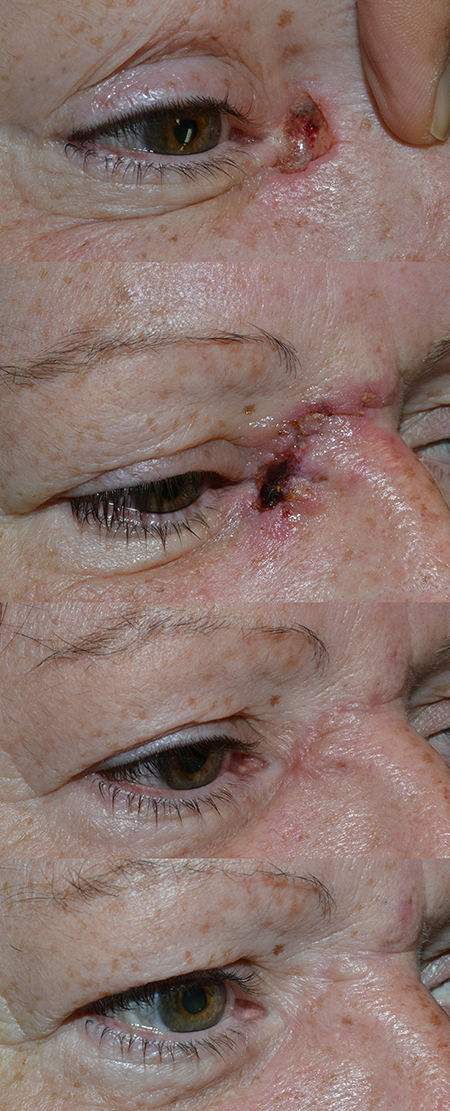
- Indications include lesion < 1.5 cm that do not involve the upper eyelid (Figure 1).
- Need to have sufficient laxity of the upper eyelid skin
- Advancement flap incision is marked in sub-brow area of upper eyelid.
- Surrounding skin is undermined, elevated, and advanced
- Combination of deep and superficial sutures placed
- A bolster is placed to allow apposition of flap to underlying tissue.
- Orbicularis oculi myocutaneous flap (Anderson, Arch Ophthalmol 1979; Reali, Ann Plast Surg 1993; Jelks, Plast Reconstr Surg 2002; d’Alcontres, Br Assoc Plast Surg 2004)
- Random skin-muscle flap of orbicularis oculi muscle and skin
- Can be advanced together, or tunneled under the skin
- Can get large areas of reconstruction (25 cm2)

Figure 1. Reconstruction of a small medial canthus defect. A. Defect after Mohs excision. B. 1 week after bilobed flap. C. 7 weeks postop with mild hypertrophy of the scar. D. 6 months postop, nearly imperceptible incision without any intervention.
- Forehead techniques
- Paramedian composite flap (Elshamma, Saudi J Ophthalmol 2013)
- Vascularized pedicle flap of skin and muscle with blood supply
- Doppler ultrasound is used to mark the course of the supraorbital or supratrochlear arteries.
- Once a sufficiently long amount is traced, the incision is marked.
- Skin and frontalis muscle is dissected from underlying galea.
- The flap is set into the medial canthal defect.
- After 6–12 weeks, the flap is divided and the edges of donor and recipient site are revised.
- Can be done with a tunneled/buried pedicle (Kim, Ann Plast Surg 2013)
- Island flaps (Motomura, 2006; Karsidag, J Craniofacial Surg 2008; Mombaerts, Dermatol Surg 2010)
- For use with large defects of the medial canthus with or without eyelid defects.
- The course of the contralateral supratrochlear artery and vein is mapped with the Doppler ultrasound.
- A skin-muscle area for reconstruction is traced.
- A subcutaneous tunnel is made under the glabella and lateral nasal wall.
- The island flap is rotated and passed through the tunnel.
- The island is sutured into place.
- Prevents the need for secondary division of pedicle.
- Forehead muscle flap (Chiarelli, Br J Plast Surg 2001)
- A vertical incision is made up the forehead.
- Subcutaneous dissection is performed.
- A random paramedian frontalis muscle flap is made, measuring 2.5 cm at the base.
- Frontalis muscle flap is raised and rotated 180 degrees to inlay into the defect.
- Split- or full-thickness skin graft is placed over the flap.
- Other techniques may also be used (tarsorrhaphy, chondro-mucosal graft).
- Closure of skin
- Pericranial flap (Leatherbarrow, Ophthal Plast Reconstr Surg 2006)
- Useful if there is loss of periosteum or bone for reconstruction
- Glabellar flap (Chao, J Plast Reconstr Aesthetic Surg 2010)
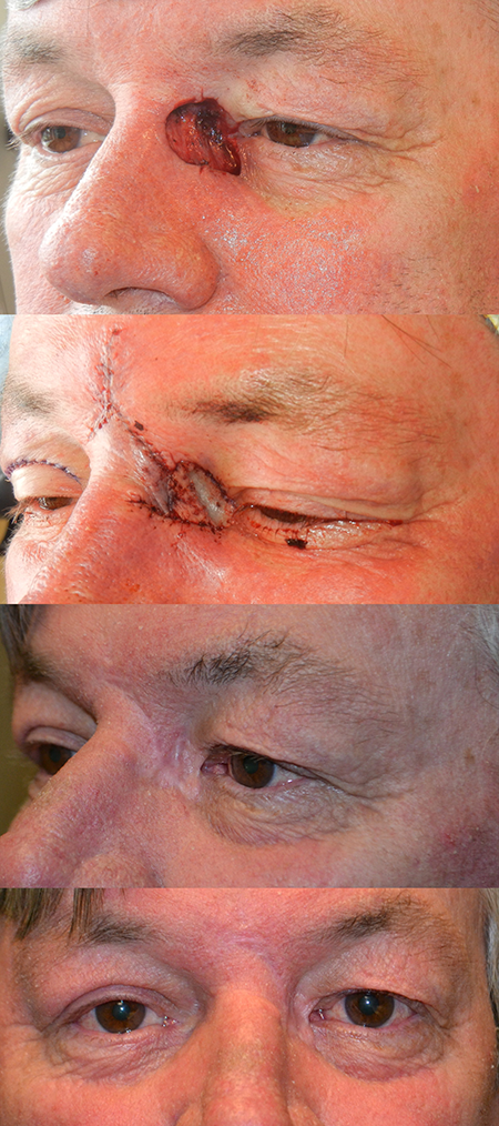
- Glabellar skin is marked in a triangular fashion, with the base wider (Figure 2).
- After incision, the skin is undermined beneath the flap and surrounding tissue.
- Glabellar flap is rotated and secured into position.
- Small dog-ear deformities at the base may need to be addressed.
- Other modifications, including the “flap-in-flap” technique, have been described (Turgut, J Craniofac Surg 2009)
- A tunneled glabellar flap can be used (Bertelmann, Ophthalmologica 2006).
- Cheek techniques
- Can use an island pedicle inferior to the defect along the nasolabial fold (Lee, Ophthal Plast Reconstr Surg 2004)
- While maintaining the pedicle base vascularity, the island is advanced superiorly into the medial canthal defect
- Donor site defect is closed horizontally.

Figure 2. Reconstruction of a moderate sized medial canthus defect. A. Moderate sized defect spanning above and below the medial canthal tendon. B. Immediate post-operative appearance of the glabellar flap with full thickness skin graft from the right upper eyelid. The thin eyelid skin graft is used to line the area immediately along the eyelid margins to allow for unrestricted movement of the eyelids. C. 9 months postop with well healed area and whitening along the suture lines. D. Note the asymmetry of eyelid creases due to the skin graft harvest.
- Graft materials
- Amniotic membrane (Harris, Ophthal Plast Reconstr Surg 2011)
- Can be used as a posterior lamellar material
- Full-thickness skin graft
- Preferable tissue for grafts
- Skin of similar texture, color, and thickness is ideal
- Must have a robust vascular bed
- Often use upper eyelid, preauricular, postauricular, or supraclavicular donor sites
- Split-thickness skin graft
- Generally avoid these grafts due to more contraction.
- Medial canthal skin is typically much thicker than split-thickness skin grafts (STSG), and therefore defects are not adequately covered
- Can be used in some instances for a very large defect
- May be considered for defect sites with vascular compromise, for example, history of radiation
- Medial canthal fixation techniques for medial canthal tendon repair
- Combined miniplate and wire (Howard, Arch Ophthalmol 1992; Wittkampf, Int J Oral Max Facial Surg 2001)
- Incision made in the medial canthus of the involved side
- Identification of the medial canthal tendon
- Place 2-0 wire through the tendon via a double stitch
- The wire is passed through the most posterior hole of a miniplate
- The anterior portion of the miniplate is fixated to the stable nasal bone in order to position the wire sufficiently posterior and superior
- The posterior end of the miniplate may need to be cantilevered into a defect for adequate posterior positioning of the tendon
- Precludes need for contralateral incision
- Micro-anchor devices, such as Mitek anchor system (Goldenberg, Ann Plast Surg 2008)
- Titanium micro-anchor device (1.3 x 3.7 mm) with prethreaded 4-0 Ethibond suture (other sizes of suture and device available)
- After exposure of the posterior lacrimal crest, a proprietary hand-powered drill is used.
- Place at level of posterior lacrimal crest and slightly superior to anterior lacrimal crest
- The screw with pre-placed suture is placed into the drill hole with pressure, and countersunk past the bone level slightly.
- Secure the tendon with the attached suture to the anchor
- Transnasal wiring (Kelly, Ophthal Plast Reconstr Surg 2004)
- A preoperative CT scan is essential to assess for a low-lying cribriform plate.
- With this technique, a skin incision is made in the medial canthi bilaterally in a Y fashion.
- After dissection is made down to periostium, periosteum is elevated in conjunction with the lacrimal sacs.
- A small drill bit is used to drill from one posterior lacrimal crest to the other.
- A 2-0 Kirschner wire is passed within the ethmoid sinuses.
- Can be performed with assistance of an 18-gauge needle
- The wire is passed through the medial canthal tendon, twisted, and pulled back to the site of insertion.
- By sufficient tightening of the wire, the two medial canthal tendons are drawn together.
Lateral canthal defects
- Eyelid techniques
- Advancement of the eyelids to the canthus (Steinkogler, Br J Ophthalmol 1983; Fuente del Campo, Ophthal Plast Reconstr Surg 1994)
- Markings are made in the lateral upper and lower eyelids along the relaxed skin tension lines.
- Dissection is carried out subcutaneously.
- The lateral upper and lower eyelids are sutured together to form a sharp lateral canthal angle.
- The lateral canthus is sutured to the lateral orbital rim.
- Skin is closed.
- A combination of techniques is often necessary (Meena, Oman J Ophthalmol 2012)
- Lateral techniques
- Semicircular flap (Tenzel, Am Acad Ophthalmol Otolargol 1978)
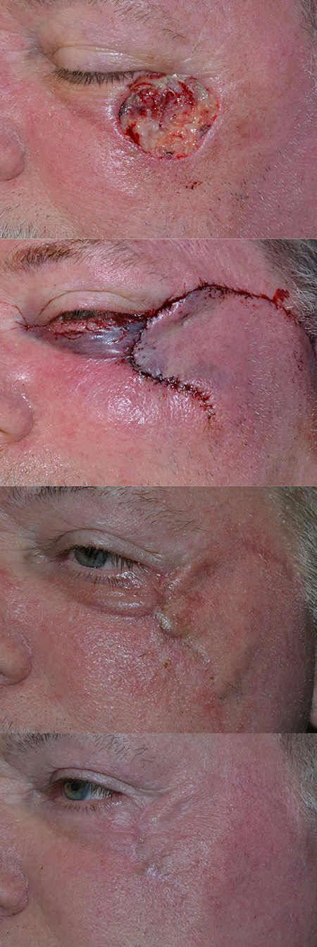
- A semicircle is drawn lateral to the lateral canthal angle (Figure 3).
- A lower eyelid defect is corrected with an inferiorly based flap; an upper eyelid defect is corrected with a superiorly based flap.
- The chord length of the semicircle should be less than 2cm in order to avoid iatrogenic injury to the facial nerve branch as it crosses over the zygomatic bone
- Subcutaneous dissection is performed under the semicircle
- The bony orbitomalar attachments to the inferolateral orbital rim may be released for mobilization of the flap
- The flap is advanced medially to the canthus to reconstruct the defect
- The base of the advanced flap should be secured to the lateral periosteum prior to completing the eyelid margin closure
- Bilobed flap (Yazici, Ophthal Plast Reconstr Surg 2013)
- A large bilobed skin flap is drawn on the lateral cheek with the base directed superiorly.
- A skin-muscle flap is dissected with undermining of the surrounding tissues to assist with closure of the defects.
- After reconstruction of the posterior lamella, the flap is inset into the lateral eyelids and canthus.
- Trilobed flap (Copcu, Plast Reconstr Surg 2006)
- Similar to the bilobed flap, but useful for upper- and lower-eyelid lesion.

Figure 3. Large lateral canthal defect. A. Size of defect immediately after Mohs excision. There was poor lateral canthal support which was bolstered with a periosteal flap. B. Immediate post-operative appearance. A rotational flap was undermined involving the majority of the lateral cheek. A subciliary incision was made as well. C. 7 weeks postop with hypertrophy of the incision at the site of the defect. Mild edema of the lateral lower eyelid. D. 6 months postop, improved eyelid position and incisional thickness without any intervention.
- Techniques using tissue from a distant location
- Island flaps
- Similar to medial canthus; however, a pedicled flap of the superficial temporal artery is used.
- Risk of injury to the temporal branch of the facial nerve.
- Graft materials
- Cadaveric acellular dermis (AlloDerm) (Dailey, Ophthal Plast Reconstr Surg 2011)
- Can be used to “bolster” the strength of lateral canthus
- After making a sideways Y incision in the lateral canthus with the arms extending to the upper and lower eyelids, dissection is carried to the tendon.
- The tendon is affixed to the periosteum of the lateral orbital rim.
- The material is sutured overlying this.
- Skin is closed.
- Porcine acellular dermis (Enduragen) (McCord, Plast Reconstr Surg 2008)
- Dermal collagen matrix that is acellular, pre-hydrated and flexible for natural contours
- Used in a similar manner for posterior lamellar support in lateral canthal reconstruction
- Two thicknesses are available and presoaking of the material prior to use is not necessary
- Lateral canthal fixation techniques
- Numerous techniques available (Chong, Facial Plast Surg 2010)
- “Webster” suture (Webster, Arch Otolaryngol 1979)
- Suture is passed through the lateral lower eyelid tarsus and periosteum of the lateral orbital rim.
- The eyelid is pulled laterally and superiorly.
- Lateral retinacular suspension (Fagien, Plast Reconstr Surg 1999)
- Suture is passed through the lateral canthal tendon and suspended superiorly up through an upper eyelid blepharoplasty incision.
- Periosteal flap (Leone, Am J Ophthalmol 1992)
- After exposure of the lateral orbital rim, a “tongue” of periosteum is incised in the lateral portion of the rim.
- The length of this “tongue” should be the distance remaining between the lateral end of the eyelid to the lateral orbital rim.
- The “tongue” is elevated with the base of the flap attached at the lateral orbital rim.
- The lateral eyelid is sutured to the flap.
- Drill hole suspension (Oh, Ophthal Plast Reconstr Surg 2013)
- Single- or double-drill hole is placed into the lateral orbital rim.
- Suture is passed through the lateral canthal tendon and through the drill hole.
- Suborbicularis oculi fat pad (SOOF) may also be suspended similarly.
- Micro-anchor devices, such as Mitek anchor system (Alfano, J Oral Maxillofac Surg 2011)
- Similar to medial canthal suspension
Instrumentation, anesthesia, and technique
- Standard surgical instruments for eyelid and facial surgery
- Local, IV sedation, or general anesthesia
Preventing and managing treatment complications
Intraoperative
- Hemorrhage
- Inadequate soft tissues surrounding the defect requires transposition flaps from the forehead or other distant tissue areas.
- Inadequate attachment to posterior lacrimal crest causes anterior displacement of the eyelid from globe, resulting in medial eyelid ectropion or punctal ectropion.
- Injury to the lacrimal collecting system
- Reconstructed medial eyelid may not have normal pump function
Postoperative
- Infection
- Epiphora
- Poor tissue approximation with eyelid margin irregularity
- Dehiscence of wound if closed under tension
- Corneal surface problems related to irregular eyelid margin and medial canthal soft tissues with loss of tear film integrity
- Ptosis of upper eyelid following repair, most often secondary to postoperative edema and horizontal tightness and will typically resolve
- Skin graft/flap necrosis: Debride as necessary, use antibiotics if infected (Figure 4)
- Hypertrophic scar formation: May be improved with injection of triamcinolone or 5-fluorouracil

Figure 4. Large lateral canthal defect with graft necrosis. A. Defect after Mohs excision. B. Immediate postoperative appearance. After relaxing incisions and undermining of adjacent tissue, the size of the defect was minimized. There was moderate canthal support which was aided by suture canthopexy. A full thickness skin graft from the supraclavicular area was placed. C. 1 month postop with early necrosis. There is elevation of the skin graft with central necrosis. This area was debrided and injected with triamcinolone. D. 2 months postop with mild scarring in area of previous skin graft.
Follow-up care
- Topical antibiotics/lubricants
- Ocular evaluation with corneal and tear film inspection
- Suture removal
- Regular dermatologic follow up
Patient Instructions
- Avoid strenuous activity or exercise for the first 1-2 weeks after surgery.
- Elevate the head of the bed while sleeping.
- Avoid eyelid trauma to the reconstructed area.
- Protective eye shield may be helpful in the first several weeks, particularly while sleeping.
- Watch for tumor recurrence.
References and additional resources
- AAO, Basic and Clinical Science Course. Section 7: Orbit, Eyelids, and Lacrimal System, 2013-2014.
- AAO, Surgery of the Eyelid, Orbit & Lacrimal system, Vol. 1, 1993, p.182-185.
- AAO, Surgery of the Eyelid, Orbit & Lacrimal system, Vol. 2, 1994, p.236-240,274-276.
- AAO, Focal Points: Management of Eyelid Trauma, Module #10, 1996.
- Alfano C, Chiummariello S, Monarca C, et al. Lateral canthoplasty by the micro-Mitek anchor system: 10-year review of 96 patients. J Oral Maxillofac Surg 2011;69:1745-9.
- Anderson RL, Edwards JJ. Reconstruction by myocutaneous eyelid flaps. Arch Ophthalmol 1979;97:2358-62.
- Behroozan DS, Goldberg LH. Upper eyelid rotation flap for reconstruction of medial canthal defects. J Am Acad Dermatol 2005;53(4):635-8.
- Bertelmann E, Rieck P, Guthoff R. Medial canthal reconstruction by a modified glabellar flap. Ophthalmologica 2006;220:368-81.
- ChaoY, Xin X, Jiangping C. Medial canthal reconstruction with combined glabellar and orbicularis oculi myocutaneous advancement flaps. J Plast Reconstr Aesthetic Surg 2010;63:1624-8.
- Chiarelli A, Forcignano R, Boatto D, Zuliani F, Bisazza S. Reconstruction of the inner canthus region with a forehead muscle flap: a report on three cases. Br J Plast Surg 2001;54:248-52.
- Chong KK, Goldberg RA. Lateral canthal surgery. Facial Plast Surg 2010;26(3):193-200.
- Copcu E, Sivrioglu N, Koc B, et al. Reconstruction of the lateral canthus with trilobed skin flap and temporalis fascia graft. Plast Reconstr Surg 2006;117(7):2514-6.
- Czyz CN, Cahill KV, Foster JA, et al. Reconstructive options for the medial canthus and eyelids following tumor excision. Saudi J Ophthalmol 2011;25:67-74.
- D’Alcontres FS, D’Amico E, Colonna MR, et al. Orbicularis oculi myocutaneous flap in the repair of the medial canthal region. A new strategy for canthal resurfacing. Br Assoc Plast Surgeons 2004;57:540-2.
- Dailey RA, Chavez MR. Lateral canthoplasty with acellular cadaveric dermal matrix graft (AlloDerm) reinforcement. Ophthal Plast Reconstr Surg 2011;28(1):e29-e31.
- Elshamma NA, Al Qabbani A, Alkatan HM, et al. The use of forehead flaps in the management of large basal cell carcinomas of the medial canthus/medial lower eyelid in Saudi patients. Saudi J Ophthalmol 2013;27:223-5.
- Emsen IM, Benlier E. The use of the superthinned inferior pedicled glabellar flap in reconstruction of small to large medial canthal defect. J Craniofac Surg 2008;19:500-4.
- Fagien S. Algorithm for canthoplasty: the lateral retinacular suspension: a simplified suture canthopexy. Plast Reconstr Surg 1999;103:2042-53.
- Fox SA, Beard C. Spontaneous lid repair. Am J Ophthalmol 1964;58:947-52.
- Fuente del Campo A, Nahas R, Vazquez Ambriz V. Procedure for the reconstruction of the lateral palpebral canthus. Ophthal Plast Reconstr Surg 1994;10(1):6-10.
- Goldenberg DC, Bastos EO, Alonso N, et al. The role of micro-anchor devices in medial canthopexy. Ann Plast Surg 2008;61:47-51.
- Harrington JN. Reconstruction of the medial canthus by spontaneous granulation (laissez-faire): a review. Ann Ophthalmol 1982;14:956-70.
- Harris MA, Vidor IA, Sivak-Calcott J. Amniotic membrane in medial canthal reconstruction. Ophthal Plast Reconstr Surg 2011;27:135-6.
- Howard GR, Nerad JA, Kersten RC. Medial canthoplasty with microplate fixation. Arch Ophthalmol 1992;110:1793-7.
- Jelks GW, Glat PM, Jelks EB, et al. Medial canthal reconstruction using a medially based upper eyelid myocutaneous flap. Plast Reconstr Surg 2002;110:1636-43.
- Karsidag S, Sacak B, Bayraktaroglu S, et al. A novel approach for the reconstruction of medial canthal and nasal dorsal defects: frontal hairline island flap. J Craniofacial Surg 2008;19(6):1653-7.
- Kelly CP, Cohen AJ, Yavuzer R, et al. Medial canthopexy: a proven technique. Ophthal Plast Reconstr Surg 2004:20(5):337-41.
- Kim JH, Kim JM, Park JW, et al. Reconstruction of the medial canthus using an ipsilateral paramedian forehead flap. Ann Plast Surg 2013;40:742-7.
- Leatherbarrow B, Watson A, Wilcsek G. Use of the pericranial flap in medial canthal reconstruction: another application for this versatile flap. Ophthal Plast Reconstr Surg 2006;22:414-9.
- Lee BJ, Elner SG, Douglas RS, et al. Island pedicle and horizontal advancement cheek flap for medial canthal reconstruction. Ophthal Plast Reconstr Surg 2011;27:376-9.
- Leone CR. Periosteal flap for lower eyelid reconstruction. Am J Ophthalmol 1992;114(4):513-4.
- Lowry JC, Bartley GB, Garrity JA. The role of second-intention healing in periocular reconstruction. Ophthal Plast Reconstr Surg 1997;13:174-88.
- McCord C, Nahai FR, Codner MA, et al. Use of porcine acellular dermal matrix (Enduragen) grafts in eyelids: a review of 69 patients and 129 eyelids. Plast Reconstr Surg 2008;122(4):1206-13.
- Meadows AER, Manners RM. A simple modification of the glabellar flap in medial canthal reconstruction. Ophthal Plast Reconstr Surg 2003;19:313-5.
- Meena M. Triple-flaps for lateral canthus reconstruction: a novel technique. Oman J Ophthalmol 2012;5(3):181-3.
- Mehta JS, Olver JM. Infraglabellar transnasal bilobed flap in the reconstruction of medial canthal defects. Arch Ophthalmol 2006;124:111-5.
- Mombaerts I, Gillis A. The tunneled forehead flap in medial canthal and eyelid reconstruction. Dermatol Surg 2010;36(7):1118-25.
- Moscona R, Pnini A, Hirshowitz B. In favor of healing by secondary intention after excision of medial canthal basal cell carcinoma. Plast Reconstr Surg 1983;71:189-95.
- Motomura H, Taniguchi T, Harada T, Muraoka M. A combined flap reconstruction for full-thickness defects of the medial canthal revion. J Plast Reconstr Aesthetic Surg 2006;59:747-51.
- Ng SGJ, Inkster CF, Leatherbarrow B. The rhomboid flap in medial canthal reconstruction. Br J Ophthalmol 2001;85:556-9.
- Oh SR, Korn BS, Kikkawa DO. Orbitomalar suspension with single drill hole canthoplasty. Ophthal Plast Reconstr Surg 2013;29(5):357-60.
- Onishi K, Maruyama Y, Okada E, et al. Medial canthal reconstruction with glabellar combined rintala flaps. Plast Reconstr Surg 2007;119:537-41.
- Reali UM, Chiarugi C, Borgognoni L. Reconstruction of a medial canthus defect with a myocutaneous flap. Ann Plast Surg 1993;30(2):159-62.
- Shankar J, Nair RG, Sullivan SC. Management of periocular skin tumors by laissez-faire technique: analysis of functional and cosmetic results. Eye 2002;16:50-3.
- Stasior OG. Reconstruction of the upper eyelid and medial canthus. Trans New Orleans Acad Ophthalmol 1982;30:392-409.
- Steinkogler FJ. Reconstruction of the temporal canthus. Br J Ophthalmol 1983;67:267-9.
- Tenzel RR, Boynton JR, Buffam FV. Technique of combined lid and medial canthus reconstruction. Ophthal Surg 1976;7(3):25-8.
- Tenzel RR, Stewart WB. Eyelid reconstruction by the semicircle flap technique. Am Acad Ophthalmol Otolargol 1978;85:1164-9.
- Tezel E, Sonmez A, Numanoglu A. Medial pedicled orbicularis oculi flap. Ann Plast Surg 2002;49:599-603.
- Turgut G, Ozcan A, Yesiloglu N, et al. A new glabellar flap modification for the reconstruction of medial canthal and nasal dorsal defects: “flap in flap” technique. J Craniofac Surg 2009;20(1):198-200.
- Webster RC, Davidson TM, Reardon EJ, et al. Suspending sutures in blepharoplasty. Arch Otolaryngol 1979;105:601-4.
- Wittkampf ARM, Mouritz MP. A simple method for medial canthal reconstruction. Int J Oral Max Facial Surg 2001;30:342-3.
- Yazici B, Cetinkaya A, Cakirlu E. Bilobed flap in the reconstruction of inferior lateral periorbital defects. Ophthal Plast Reconstr Surg 2013;29:208-14.
- Yildirim S, Akoz T, Akan M, et al. The use of combined nasolabial V-Y advancement and glabellar flaps for large medial canthal defects. Dermatol Surg 2001;27:215-8.
