Milia
Updated Nov. 2024
Alexis Kassotis and Lora R. Dagi Glass, MD
Establishing the diagnosis
Numerous forms of milia exist, with the ones most likely to involve the periocular area discussed in this review.
Etiology
- Milia are miniature, superficial, keratinous cystic lesions.
- Primary milia (i.e. milia that occur independent of underlying disease) are thought to arise from sebaceous ducts surrounding vellus hairs.
- Secondary milia (i.e. milia that occur in the setting of another medical condition) are thought to arise from eccrine sweat ducts.
- Milia en plaque (MEP) is characterized by the development of numerous milia on erythematous plaques.
- MEP can occur as a form of primary milia or as a form of secondary milia.
- Secondary MEP is usually reported in the setting of discoid lupus erythematosus, lichen planus, pseudoxanthoma elasticum, or cyclosporine use.
- MEP can occur as a form of primary milia or as a form of secondary milia.
- Multiple eruptive milia (MEM) is characterized by the spontaneous eruption of a significantly larger number of milia than is seen in other forms.
- Different forms of MEM have been identified, with various primary and secondary etiologies:
- Isolated idiopathic
- Familial autosomal dominant
- One reported variant of MEM is characterized by milia, isolated to the eyelids, occurring in one family over multiple generations (Ratnavel, 1995).
- Associated with genodermatoses (a diverse group of inherited conditions with cutaneous manifestations).
- Different forms of MEM have been identified, with various primary and secondary etiologies:
- There is evidence that milia can arise in areas of previous epithelial injury.
- Milia has been reported to erupt within a tattoo and in the scar of a herpes zoster lesion (Ross, 2017 & Lee, 1996).
Epidemiology
- Primary milia
- Congenital milia
- Occurs in up to 50% of newborns (Berk, 2008).
- Less common in premature newborns.
- Benign primary milia:
- Common; develops spontaneously in children and adults.
- Congenital milia
- Secondary milia
- Feature of multiple genodermatoses:
- Some examples include:
- Nevoid basal cell carcinoma syndrome
- Specifically associated with periocular milia, which occurs in approximately 30% of cases (Berk, 2008).
- Epidermolysis bullosa
- Porphyrias
- A sporadic case of a Bazex-Dupre-Christol-like syndrome, which leads to early onset basal cell carcinoma, presented with prominent periocular milia, hypohidrosis and hypotrichosis (Glaessl, 2000).
- Nevoid basal cell carcinoma syndrome
- Some examples include:
- Feature of multiple genodermatoses:
- MEP is a rare condition.
- Fewer than 30 cases have been reported (Berk, 2008).
- Most cases occur in middle aged females.
- Most cases present in the periauricular region, but MEP of the eyelids has been reported (Bridges, 2009 & Wong, 1999).
- Fewer than 30 cases have been reported (Berk, 2008).
- MEM is also a rare condition.
- It is most commonly associated with genodermatoses.
- Few isolated cases have been reported, affecting individuals 15 to 71 years of age.
Clinical features
- Congenital milia and benign primary milia
- 1-3mm, round, white papules.
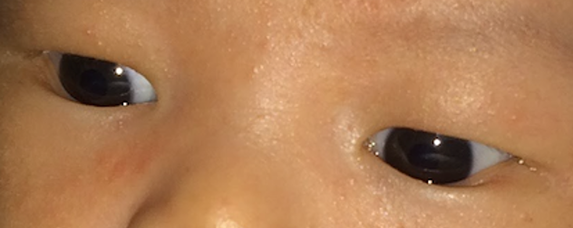
- Often involving the forehead, cheeks and eyelids (Figure 1).
- Benign primary milia usually persists for weeks to months longer than congenital milia.
- MEP
- Erythematous, indurated plaques, several centimeters in diameter, covered with numerous milia.
- Classically erupts on the head and neck.
- MEM
- Continuous eruption of a large number of milia for weeks to months.
- Classically erupts on the head.
Other diagnostic studies
- Wood’s lamp
- Milia fluoresce bright yellow.
- This may be used if diagnosis in uncertain.
- Workup for underlying disease in the appropriate clinical situation (i.e. abrupt onset of periocular milia in a young adult may warrant thorough skin examination for basal cell carcinoma).
Differential diagnosis
- Congenital milia and benign primary milia
- Sebaceous hyperplasia
- Neonatal cephalic pustulosis
- Miliaria
- Epidermal inclusion cyst
- Molloscum contagiosum
- MEP
- Follicular mucinosis
- Trichoadenoma
- Tumidus follicularis
- MEM
- Benign primary milia
Patient management
- Medical therapy: in most cases, treatment is not required.
- Topical retinoids have been shown to be rapidly efficacious for eruptions of multiple lesions (i.e. extensive congenital or benign primary milia and MEM).
- Surgical therapy: if desired for cosmetic purposes.
- Incision of the epidermis with expression of keratinaceous material is used for single or few lesions (i.e. nick skin with 11 blade or bent needle and express using gentle pressure at the base with forceps).
- Electrodessication has been used successfully to treat periocular MEP (Al-Mutairi, 2006).
- Mild electrocautery has also been used successfully for treatment of numerous milia.
- Laser ablation using CO2 fractional laser (Tenna, 2014) and erbium:YAG (Voth, 2011) has also demonstrated efficacy in treating periocaular MEP.
- Prognosis: self-limiting
- Resolves within weeks to months.
- Recurrence is rare.
Disease-related complications
- None directly related to milia; if milia arise in the setting of an underlying condition, such as described above, complications of that condition may occur.

Photograph courtesy of Michael Yoon, MD.
Figure 1. Congenital milia involving the eyelids bilaterally.
References and additional resources
- Berk D & Bayliss S. Milia: A review and classification. J Am Acad Dermatol. 2008;59(6):1050-1063.
- Connelly T. Eruptive milia and rapid response to topical tretinoin. Arch Dermatol. 2008;144(6):816-817.
- Lee WS, Kim SJ, Ahn SK, Lee SH. Milia arising in herpes zoster scars. J Dermatol. 1996;23(8):556-558.
- Ross N, Farber M, Sahu J. Eruptive milia within a tattoo: A case report and review of the literature. J Drugs Dermatol. 2017;16(6):621-624.
- Wong, Goh. Milia en plaque. Clin Exp Dermatol. 1999;24(3):183-185.
- Dogra S, Kaur I, Handa S. Milia en plaque in a renal transplant patient: a rare presentation. Int J Dermatol. 2002;41:897-898
- Lee JH, Kwon HS, Jung HM, et al. Wood’s lamp-induced fluorescence of milia. J Am Acad Dermatol. 2018;78(5):e99.
- Glaessl A, Hohenlautner U, Landthaler M, Vogt T. Sporadic Bazex–Dupré–Christol‐like Syndrome: Early Onset Basal Cell Carcinoma, Hypohidrosis, Hypotrichosis, and Prominent Milia. Dermatologic Surgery. 2000; 26(2):152-154.
- Ratnavel RC, Handfield-Jones SE, Norris PG. Milia restricted to the eyelids. Clin Exp Dermatol. 1995; 20:153-154.
- Cordero I. Electrosurgical units – how they work and how to use them safely. Community Eye Health. 2015;28(89):15‐16.
- Gorlin RJ. Nevoid basal-cell carcinoma syndrome. Medicine (Baltimore). 1987;66(2):98‐113.
- Al-Mutairi N & Arun J. Bilateral Extensive Periorbital Milia en Plaque Treated with Electrodesiccation. J Cutan Med Surg. 2006;10(4):193-6.
- Tenna S, Filoni A, Pagliarello C, Paradisi M, Persichetti P. Eyelid milia en plaque: a treatment challenge with a new CO2 fractional laser. Dermatol Ther. 2014;27(2):65-67.
- Voth H, Reinhard G. Periocular milia en plaque successfully treated by erbium:YAG laser ablation. J Cosmet Laser Ther. 2011;13(1):35-37.
