Bacterial Orbital Cellulitis
Updated May 2024
Establishing the diagnosis
Etiology
- Identify etiology to determine likely microbiology and appropriate therapy.
- Balance between flora and defense mechanisms
- Millions of microbes surround the orbit, e.g., on the eyelid skin/adjacent sinuses.
- Natural host defenses, e.g., immune system/anatomic barriers such as the orbital septum help to maintain a microbe-free environment within the orbit.
- Orbital cellulitis can occur if there is a weakening or breach of these normal host defenses or alternatively in cases of microbial hypervirulence (Liao, Ophthalmology 2015).
Normal defenses
Hypervirulent organisms, e.g., MRSA, can pass posteriorly through a normal orbital septum.
Compromised defenses
- Sinusitis-related orbital cellulitis (primary cause of orbital cellulitis)
- In sinusitis, sinus obstruction results from allergic rhinitis or viral inflammation:
- Bacterial proliferation results from these obstructions, and their inflammatory products potentiate it (Harris, Ophthalmology 1994).
- Age-related sinus anatomic differences help to determine local conditions (pO2, pCO2, pH) that allow aerobic and/or anaerobic microbes to flourish (Harris, Ophthalmology 1994).
- With increasing age, the sinus cavities enlarge markedly, but the sinus ostia remain approximately the same size.
- Relative width of sinus ostia in young children partially explains their greater incidence of acute sinusitis, since frequent upper respiratory infections tend to involve the nose and sinuses as a single structure.
- Consequently, younger children rarely achieve strict anaerobic conditions in their sinuses, while older children and adults are prone to more complete sequestration and complex infections (Harris, Ophthalmology 1994).
- Very thin bone separates the adjacent sinuses from the orbit, e.g., ethmoid bone, allowing direct passage of bacteria through small bony dehicenses, e.g., vascular canals.
- Orbital complications of sinusitis in the first decade typically result from disease in the ethmoid and maxillary sinus (Harris, Ophthalmology 1994); The frontal sinus first appears between ages 5 and 7 years, but does not fully develop until late adolescence
- Breach/bypass of normal orbital barriers
- Trauma
- Common organisms: Staphylococcus species, Streptococcus species, gram-negative rods
- Fracture: Orbital cellulitis following orbital fracture is rare, and more common when paranasal sinus infection preexists or occurs within several weeks of injury (Ben Simon, Ophthalmology 2005).
- Eyelid laceration/cutaneous abrasion (Mills OPRS 2008)
- Orbital or sinus foreign body (Levy, OPRS 2004)
- Postsurgical
- Common organisms: Staphylococcus species, Streptococcus species
- Glaucoma surgery (La Vina, Arch Ophthalmol 2002).
- Scattered case reports
- Historically, responds to systemic antibiotics, and has not required removal of the implant
- Orbital fracture repair
- Ben Simon, et al. (2005) identified only 4 cases of orbital cellulitis out of 497 patients seen for orbital fractures.
- Wladis (2013) reported no cases of postoperative cellulitis in 153 patients who had fracture repair.
- Strabismus surgery: Estimated rate of periocular infection in 1/1,000 surgeries (Dhrami-Gavazi, OPRS, 2014)
- Retrobulbar alcohol and chlorpromazine injections (Margo, Arch Ophthalmol 1993).
- Orbital surgery
- Very little data regarding incidence
- Fay (OPRS 2013): only 2 cases of presumed cellulitis after enucleation or evisceration
- Lacrimal probing or surgery
- Sinus surgery
- Dental procedures
- Eyebrow piercing
- Endogenous spread
- Often associated with immunodeficiency
- Pseudomonas aeruginosa septicemia (Maccheron, AJO 2004)
- Klebsiella septicemia from liver abscesses (Davies, OPRS 2014)
- Adjacent tissue spread
- Dental abscess leading to odontogenic orbital cellulitis (Youssef, OPRS 2008) typically polymicrobial (anaerobic and anaerobic)
- Intracranial infection or abscess
- Endophthalmitis
- Postoperative
- Endogenous
- Primary or secondary systemic immunodeficiency/immunocompromised state
- Common organisms:
- Primarily fungal
- Mucormycosis
- Aspergillosis
- Streptococcus pneumonia
- Hemophilus influenzae
- HIV
- Diabetic ketoacidosis
- CLL/Multiple Myeloma
- Cancer chemotherapy
- Lymphoma
- Glucocorticoid therapy
Epidemiology
The Scottish Ophthalmic Surveillance Unit reported the combined incidence of 1.6 cases per 100,000 children and 0.1 cases per 100,000 adults of all forms of orbital cellulitis (Murphy, BJO 2014).
47% of children had a URI and 87% had sinus disease.
Immunosuppression and trauma were the leading causes in adults.
There was no racial or gender predilection.
Sinusitis-related orbital cellulitis is more common in winter months in northern climates due to an increased incidence of sinusitis and URI (Bergin, BJO 1986).
History
- Typically acute onset of symptoms
- Symptoms
- Malaise
- Fever
- Pain, especially with eye movements
- Vision loss
- Diplopia
- History of sinus infections, surgery or trauma
- Relevant medical history: diabetes mellitus, especially with ketoacidosis
- History of immunocompromise
Clinical features
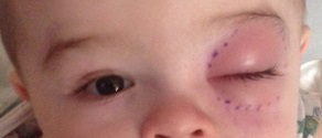
- General signs (Figure 1)
- Proptosis
- Restricted extraocular movement/Pain with eye movement
- Decreased vision
- Afferent pupillary defect
- Reduced corneal sensation
- Eyelid edema
- Erythema, tenderness, warmth
- Conjunctival injection
- Chemosis
- Fever
- Eschar on roof of mouth and/or in sinuses (mucormycosis)

Figure 1. Clinical photo displaying classic clinical signs: well-demarcated severe eyelid edema and erythema.
Signs of potential orbital subperiosteal abscess include proptosis and mass effect.
- Globe or optic nerve compression threatening vision
- Extraocular muscle compression producing diplopia
- Increased pain
Testing
Detailed clinical exam
- Vision
- Color vision
- Pupillary/motility exam
- Hertel measurements
- Anterior and posterior segment exam
Orbital imaging
- Computed tomography (CT) orbits
- Orbital fat stranding
- Orbital abscess (subperiosteal)
- Adjacent sinus disease
- Magnetic Resonance Imaging (MRI)
- Role secondary to CT
- Can be useful to determine the extent of soft tissue involvement
Laboratory studies
- Complete blood count (CBC)
- CRP levels correlate with duration of hospitalization in children with soft tissue infections (Tanir, Jpn J Infect Dis 2006)
- Basic metabolic panel
- Blood cultures
Biopsy (when clinically indicated)
- Tissue culture for fungal or atypical mycobacteria
- Biopsy of nasal/sinus mucosa or necrotic skin lesion
Testing for staging, fundamental impairment
Initial CT scans are not necessarily predictive of the clinical course in patients with a subperiosteal abscess. CT scan findings can lag behind the clinical picture and should not routinely be used for clinical monitoring (Harris OPRS 1996).
Risk factors
Risk factors include recent infection, e.g., sinus, upper respiratory, dacryocystitis, bacteremia, tooth abscess, trauma, or facial surgery.
Compromised immune function can lead to easier breach of the typically bacterial-free environment within the orbit.
Differential diagnosis
Infection
- Preseptal cellulitis
- Nonbacterial forms of orbital cellulitis, e.g., mucormycosis/aspergillosis
- Necrotizing fasciitis
- Myiasis: Larvae of flies can parasitize human tissue and cause an orbital inflammatory response.
- Oestris ovis is the most common cause of external ophthalmomyiasis in the United States (eyelids and orbit).
- Primary host is sheep.
- Dermatobia hominis is a botfly that can cause cutaneous eyelid infection and orbital cellulitis whose primary host is humans.
- Commonly associated with travel to Central and South America, but has been observed in Florida
- Cuterebra species have caused palpebral myiasis in North America (Engelbrecht, Arch Ophthalmol 1998).
- Primary hosts are rodents and rabbits.
Inflammation
- Thyroid eye disease
- Idiopathic orbital inflammation
- Granulomatosis with polyangiitis
- Sarcoidosis
- Systemic lupus erythematosis
- Endophthalmitis
- Posterior scleritis
- Ruptured dermoid cyst
- Medication-induced orbital inflammation (i.e. bisphosphonates, ipilimumab)
Trauma
- Hemorrhage
- Emphysema
Malignancy
- Lymphoproliferative disease: Natural killer/T-cell lymphoma can present as orbital cellulitis (Charlton OPRS 2008).
- Sinus tumor extension
- Metastatic tumor
- Esophagus (Oh, Arch Ophthalmol 2000)
- Breast
- Lung
- Prostate
- Retinoblastoma: Among 292 patients with retinoblastoma at King Khaled Hospital in Saudi Arabia, 14 (5%) presented with signs of cellulitis, culture negative, with necrosis and inflammation (Mullaney, BJO 1998).
- Neuroblastoma
- Plasma cell tumor can mimic bacterial orbital cellulitis presentation (Kelly, BJO 1991).
- Orbital melanoma can present as orbital cellulitis (Fezza, OPRS 1998).
- Ruptured dermoid cyst
Vascular
- Carotid cavernous fistula
- Cavernous sinus thrombosis
- Orbital venous malformation
- Superior vena cava syndrome
Patient management: treatment and follow-up
Natural history
The natural history of each case of orbital cellulitis varies based on the etiology, microbiology and immune status, e.g., course of MRSA orbital cellulitis from a skin source differs from sinusitis-related MRSA orbital cellulitis.
General considerations
Orbital cellulitis is an end organ–threatening condition and a potentially fatal illness.
With initial diagnosis, hospital admission with treatment and close observation is preferred over clinic-based management.
Consider infectious disease (ID) and ear, nose, and throat (ENT) consultation and comanagement.
Medical therapy
Normal defenses
- Hypervirulent organism, e.g., MRSA
- Clinical practice guidelines have been developed for skin and soft-tissue infections, bacteremia and endocarditis, pneumonia, bone and joint infections, and CNS infections (Stevens, Clin Infect Dis 2014).
- Recommendations do not directly involve orbital cellulitis.
- Most common initial IV antibiotics choice: Clindamycin/Vancomycin; consider dual therapy with third-generation cephalosporin.
- Consider ID consult.
Compromised defenses
- Sinusitis-related orbital cellulitis — primary cause of orbital cellulitis
- Children < 9 years old: typically single pathogen aerobic infections (Streptococcus and Staphylococcus strains)
- Children 9–15 years old: increasing prevalence of polymicrobial disease
- Adults: polymicrobial infections (+/- anaerobes)
- Dual therapy IV antibiotics: either amoxicillin-sulbactam or a third-generation cephalosporin (cover most aerobes and anaerobes) plus either clindamycin/vancomycin (MRSA coverage)
- Twice daily oxymetazoline nasal spray
- Expectant management:
- Careful monitoring for an afferent pupillary defect, at least as often as every 2 hours (nursing) and every 8 hours (house staff)
- Default to surgery if
- An APD develops at any time
- Failure to defervesce after 36 hours of appropriate ABX
- Clinical deterioration despite 48 hours treatment
- No improvement after 72 hours
- Systemic corticosteroids may shorten hospitalization and/or antibiotic duration, but this addition to the treatment regimen remains limited by low level evidence. (Yen OPRS 2005, Yoon Semin Ophthalmol 2023).
- The mean and median duration of IV antibiotics in a cohort of 42 patients with nonsurgical subperiosteal abscess management was 4 days (range 2–8 days) with post-discharge oral antibiotic treatment for 2 to 3 weeks (Emmett Hurley OPRS 2012).
- Breach/bypass of normal orbital barriers: either amoxicillin-sulbactam or a third-generation cephalosporin cover most aerobes and anaerobes +/- MRSA coverage
- Primary or secondary systemic immunodeficiency/immunocompromised state
- Either amoxicillin-sulbactam or a third-generation cephalosporin cover most aerobes and anaerobes +/- MRSA coverage
- Correct underlying immunodeficiency if possible, e.g., diabetic ketoacidosis
- Strongly consider ID consult
Surgery
Normal defenses
- Hypervirulent organism, e.g., MRSA
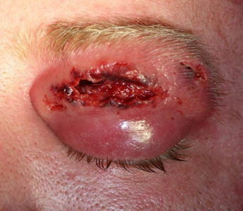
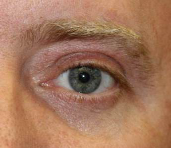
- Surgical debridement necrotic tissue should be considered (Figure 2)


Figure 2. A. Immediate postoperative photo following debridement of necrotic tissue from MRSA skin wound. No closure was performed. B. 6-month result.
Compromised defenses
- Sinusitis-related orbital cellulitis (#1 cause of orbital cellulitis)
- Subperiosteal abscess (SPA) can develop adjacent to infected sinus and is typically an extension of sinus infection through orbital wall causing subperiosteal orbital purulence without generalized orbital involvement
- SPA management (Liao, Ophthalmology 2015)
- Emergent drainage: patients of any age whose optic nerve or retinal function is compromised by the mass effect of an abscess
- Urgent drainage within 24 hours
- Large SPA
- SPAs that extend superiorly or inferiorly beyond the medial subperiosteal space
- Frontal sinus involvement
- Intracranial extension
- Presumed anaerobic infection
- Patients ≥ 9 years of age (risk for anaerobic infection)
- The emergence of aggressive aerobic pathogens, e.g. MRSA, in the context of sinusitis-related disease does not lead to modification of the above treatment algorithm
- In the setting of SPA > 2 cm with concurrent sinusitis, combined sinus drainage surgery and subperiosteal drainage has been associated with improved treatment outcome versus SPA drainage alone (Dewan, OPRS 2011)
Breach/bypass of normal orbital barriers
The sinusitis-related SPA protocol is not directly applicable to other forms of SPA, although general principals might be useful, e.g., careful monitoring for APD, failed improvement prompting surgical drainage.
Primary or secondary systemic immunodeficiency/immunocompromised state
The sinusitis-related SPA protocol is not directly applicable to other forms of SPA, although general principals might be useful, e.g., careful monitoring for APD, failed improvement prompting surgical drainage.
Common antibiotics
- Ampicillin/sulbactam (Unasyn)
- Sulbactam = beta-lactamase inhibitor
- Covers Staph, Strep, anaerobes and Enterobacteriaceae
- Does not cover pseudomonas or MRSA
- Piperacillin/tazobactam (Zosyn)
- Tazobactam = beta-lactamase inhibitor
- Spectrum similar to Unasyn, covers pseudomonas
- Does not cover MRSA
- Third-generation cephalosporin (Cefotaxime, ceftriaxone, ceftazidime)
- Covers Staph, Strep, and Enterobacteriaceae
- Does not cover anaerobes, pseudomonas, or MRSA
- MRSA coverage if suspected
- Clindamycin
- Vancomycin
Disease-related complications
- Subperiosteal/orbital abscess
- Visual loss (Arch Ophthalmol 1987; 105:345)
- Central retinal artery occlusion (OPRS 2013; 29:e59)
- Superior ophthalmic vein thrombosis (OPRS 2005; 21:387)
- Orbital compartment syndrome (OPRS 2012; 28:7)
- Cavernous sinus thrombosis (OPRS 2008; 24:408)
- Meningitis
- Brain abscess
- Cranial neuropathy
- Restrictive myopathy
- Death
Historical perspective
In 1926 a case of blindness from orbital cellulitis complicating an infected hordeolum was reported (AJO 1926; 9:34).
Gamble in 1936 reviewed a preantibiotic series of 25 children with orbital cellulitis at the Children’s Memorial Hospital in Chicago, in which 17% died of intracranial extension and 20% developed blindness (Arch Ophthalmol 1936; 16:483).
In 1948, Smith and Spencer defined groups orbital complications from sinusitis (Ann Otol Rhinol Laryngol 1948; 57:5), later modified by Chandler et al. prior to the CT era (Laryngoscope 1970; 80:1414).
Subperiostial abscess was recognized radiographically with the introduction of CT scans in the 1970s.
In 1976, Watters reviewed 10 years’ experience with intravenous methicillin and ampicillin in 104 patients with bacterial orbital cellulitis treated at the Children’s Hospital of Pittsburgh (Arch Ophthalmol 1976; 94:785).
In 1993, Harris described the importance of age as a factor in the bacteriology and response to treatment of sinusitis-associated subperiosteal abscess (Ophthalmology 1994;101:585).
References and additional resources
- http://eyewiki.org/Orbital_Cellulitis
- https://www.aao.org/theeyeshaveit/red-eye/orbital-cellulitis.cfm
- http://www.ophthalmology.theclinics.com/article/s0896-1549(05)70221-2/abstract
- AAO, Basic and Clinical Sciences Course. Section 7: Orbit, Eyelids, and Lacrimal System, 2013-2014.
- AAO, Focal Points: Preseptal and Orbital Cellulitis, Module #11, 2008.
- Ben Simon GJ, Bush S, Selva D, et al. Orbital cellulitis: a rare complications after orbital blowout fracture. Ophthalmology, 112: 2030-4, 2005.
- Bergin DJ and Wright JE. Orbital Cellulitis. Br J Ophthalmol 1986;70:174-78.
- Cannon PS, Mc Keag D, Radford R, et al. Our experience using primary oral antibiotics in the management of orbital cellulitis in a tertiary referral centre. Eye (Lond) 2009;23(3):612-5.
- Charlton J et al. Natural killer/T-cell lymphoma masquerading as orbital cellulitis. OPRS 2008; 24:143-5.
- Davies BW, Fante RG. Concurrent Endophthalmitis and Orbital Cellulitis From Metastatic Klebsiella pneumonia Liver Abscess. Ophthal Plast Reconstr Surg 2014; epub ahead of print.
- Dewan MA et al. Orbital cellulitis with subperiosteal abscess: demographics and management outcomes. OPRS 2011;27:330
- Dhrami-Gavazi E, Lee W, Garg A, et al. Bilateral orbital cellulitis after strabismus surgery. Ophthal Plast Reconstr Surg, 2014, epub ahead of print.
- Emmett Hurley P, Harris GJ. Subperiosteal abscess of the orbit: duration of intravenous antibiotic therapy in nonsurgical cases. OPRS 2012;28:22-6.
- Engelbrecht NE et al. Palpebral myiasis causing preseptal cellulitis. Arch Ophthalmol 1998; 116:684.
- Fay A, Nallassamy N, Pemberton JD, et al. Prophylactic postoperative antibiotics for enucleation and evisceration. Ophthal Plast Reconstr Surg, 29: 281-4, 2013.
- Fezza J et al. Orbital melanoma presenting as orbital cellulitis: a clinicopathologic report. OPRS 1998; 14:286
- Garcia GH, Harris GJ. Criteria for nonsurgical management of subperiosteal abscess of the orbit: analysis of outcomes 1988-1998. Ophthalmology 2000;107(8):1454-6; discussion 7-8.
- Goldstein SM, Shelsta HN. Community-acquired Methicillin-resistant Staphylococcus aureus Periorbital Cellulitis: A Problem Here to Stay. Ophthal Plast Reconstr Surg 2009;25(1):77.
- Harris GJ. Subperiosteal abscess of the orbit. Age as a factor in the bacteriology and response to treatment. Ophthalmology 1994;101(3):585-95.
- Harris GJ. Subperiosteal abscess of the orbit: computed tomography and the clinical course. Ophthal Plast Reconstr Surg 1996;12(1):1-8.
- Hurley E, Harris GJ. Subperiosteal abscess of the orbit: duration of intravenous antibiotic therapy in nonsurgical cases. Ophthal Plast Reconstr Surg 2012;28(1):22-6.
- Kelly SP et al. Solitary extramedullary plasmacytoma of the maxillary antrum and orbit presenting as acute bacterial orbital cellulitis. BJO 1991; 75:438-9.
- Kloek CE, Rubin PA. Role of inflammation in orbital cellulitis. Int Ophthalmol Clin 2006;46:57-68.
- Kronish JW, Johnson TE, Gilberg SM, Corrent GF,McLeisch WM, Scott KR. Orbital infections in patients with human immunodeficiency virus infection. Ophthalmology 1996;103(9):1483-92.
- La Vina AM et al. Orbital cellulitis as a late complication of glaucoma shunt implantation. Arch Ophthalmol 2002; 120:849-51.
- Levy J et al. Intrasinus wood foreign body causing orbital cellulitis in congenital insensitivity to pain with anhidrosis syndrome. OPRS 2004; 20:81-3.
- Liao JC, Harris GJ. Subperiosteal abscess of the orbit: evolving pathogens and the therapeutic protocol. Ophthalmology 2015;122(3):639-47.
- Lu JE, Yoon MK. The Role of Steroids for Pediatric Orbital Cellulitis – Review of the Controversy. Semin Ophthalmol. 2023 Jul;38(5):442-445. doi: 10.1080/08820538.2023.2168487. Epub 2023 Jan 22. PMID: 36683269.
- Maccheron LJ et al. Orbital cellulitis, panophthalmitis, and ecthyma gangrenosum in an immunocompromised host with pseudomonas septicemia. AJO 2004;127:176-8.
- Margo CE, Wilson T. Orbital cellulitis after retrobulbar injection of chlorpromazine. Arch Ophthalmol 1993; 111:1322.
- Marcet MM, Woog JJ, Bellows RA, et al. Orbital complications after glaucoma drainage device procedures. Ophthal Plast Reconstr Surg, 21: 67-9, 2005.
- McKinley SH, Yen MT, Miller AM, Yen KG. Microbiology of pediatric orbital cellulitis. Am J Ophthalmol 2007;144(4):497-501.
- McKinley SH, Yen MT, Miller AM, Yen KG. Microbiology of pediatric orbital cellulitis. Am J Ophthalmol 2007;144(4):497-501.
- Miller A, Castanes M, Yen M, et al. Infantile orbital cellulitis. Ophthalmology 2008;115(3):594.
- Mills DM, Meyer DR. Posttraumatic cellulitis and ulcerative conjunctivitis caused by Yersinia enterocolitica O:8. OPRS 2008; 24:425-6.
- Mullaney PB et al. Retinoblastoma associated orbital cellulitis. BJO 1998; 82:517-21.
- Murphy C et al. Orbital cellulitis in Scotland: current incidence, aetiology, management and outcomes. BJO 2014;98:1575-8.
- Nageswaran S, Woods CR, Benjamin DK, Jr., et al. Orbital cellulitis in children. Pediatr Infect Dis J 2006;25(8):695-9.
- Oh KT et al. Adenocarcinoma of the esophagus presenting as orbital cellulitis. Arch Ophthalmol 2000; 118:986-8.
- Papavasileiou E, Prasad S, Freitag SK, Sobrin L, Lobo AM. Ipilimumab-induced Ocular and Orbital Inflammation–A Case Series and Review of the Literature. Ocul Immunol Inflamm. 2016;24(2):140-6. doi: 10.3109/09273948.2014.1001858. Epub 2015 Mar 11. PMID: 25760920.
- Pirbhai A, Rajak SN, Goold LA, Cunneen TS, Wilcsek G, Martin P, Leibovitch I, Selva D. Bisphosphonate-Induced Orbital Inflammation: A Case Series and Review. Orbit. 2015;34(6):331-5. doi: 10.3109/01676830.2015.1078380. Epub 2015 Nov 5. PMID: 26540241.
- Stevens DL et al. Avoiding the perfect storm: the biologic and clinical case for reevaluating the 7-day expectation for methicillin-resistant Staphylococcus aureus bacteremia before switching therapy. Clin Infect Dis 2014; 59:e10-52
- Tanir G, Tonbul A, Tuygun N, et al. Soft tissue infections in children:a retrospective analysis of 242 hospitalized patients. Jpn J Infect Dis, 59:258–60, 2006.
- Wladis EJ. Are post-operative oral antibiotics required after orbital floor fracture repair? Orbit, 32: 30-2, 2013.
- Yen MT, Yen KG. Effect of corticosteroids in the acute management of pediatric orbital cellulitis with subperiosteal abscess. OPRS 2005; 21:363-7.
- Youssef OH, Stefanyszyn MA, Bilyk JR: Odontogenic orbital cellulitis. OPRS 2008; 1:29.
