Periocular Burns
Updated May 2024
Establishing the diagnosis
Etiology
Hot-water scalds and other thermal injuries in the home are the most common causes of periocular burns.
Flame and flash flame injuries in the workplace are also relatively frequent injuries.
Chemical-related burns, such as from acid and alkali substances, can also cause soft tissue periocular injury, but are more commonly associated with direct ocular injury.
Epidemiology
Every year in the US, there are around 450,000 burn-related healthcare visits and around 40,000 hospitalizations.
Around 3/4ths of hospitalizations are at burn centers.
More than 3% of burn-center patients do not survive.
Periocular burns are found in 50%–85% of patients admitted to burn units.
Ocular injury is reported in 7.5%–30% of burn-unit patients, with the most common injury being corneal epithelial defects and conjunctival injury.
More than 60% of lid injuries are bilateral.
History
- Timing of injury
- Mechanism of injury (explosive injury should prompt clinical and radiologic evaluation for foreign body)
- History of contact lens wear (require removal)
- History of treatments that would affect exam (fluid resuscitation, pharmacologic therapy that would induce pupillary changes)
- History of immunocompromise
- History of thermal injury in enclosed space (risk for inhalation injury)
Clinical features
- Clinical features of skin damage from burns varies depending on the timing of the exam and depth of the burn:
- Early skin damage spans from:
- Blanching erythema with mild pain
- Blistered erythema with severe pain
- White featureless thickened skin with areas of numbness
- Soft necrotic skin with exposed deep tissues
- Late skin findings include edema and tissue whitening and progression to ischemia and eschar with possible superinfection.
- Eyelid-specific burn features can include
- Loss of eyebrow and eyelash hairs
- Concomitant lacerations
- Eyelid avulsions with canalicular lacerations
- Orbital compartment syndrome with elevated IOP (from injury or resuscitation-related edema)
- Ocular injuries include
- Corneal epithelial defects
- Conjunctival chemosis
- Corneal thinning, melting or perforation
- Conjunctival tissue necrosis
- Cataract (from flash/arc burns)
- Foreign bodies (from the injury or from related field interventions)
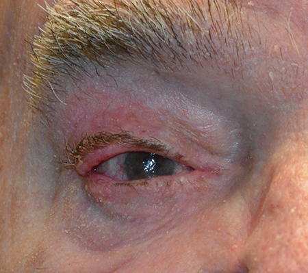
- Contractures and eyelid malposition occur as early as days after injury, but most often around 2 weeks (Figure 1).

Figure 1. Patient 3 weeks after a deep dermal burn to the right upper eyelid with early anterior lamella contraction and upper eyelid ectropion/distraction.
- Capillary leak related to systemic inflammatory response in burn fields that are greater than 20% of total body surface area require fluid resuscitation. The head and face compose 9% of total body surface area.
- Burn wound anemia refers to anemia after severe burns caused by a decrease in the half-life of circulating red blood cells.
Testing
- Early evaluation
- Evaluate skin and outline area of burn
- Evaluate Bell’s phenomenon
- Evaluate for eyelash loss to predict margin involvement
- Slit lamp exam for corneal epithelium and stromal damage or stromal thinning
- Evaluate intraocular pressure
- Consider CT to evaluate for foreign bodies
- Sedated patients, patients undergoing large-volume fluid resuscitation, and patients on positive pressure ventilation may have edema and effort-related lagophthalmos independent of eyelid burns.
- Late evaluation
- Evaluation for malposition of eyelids and lagophthalmos
Testing for staging, fundamental impairment
Skin burn damage features can be predicted and classified by the depth of injury:
- First-degree burns (epidermal burns): Erythematous, unblistered skin changes with vasodilation and minimal edema.
- Second-degree burns (partial thickness burns involving epidermis and dermis)
- Dermal burns result in vasodilation, edema and inflammation.
- Superficial dermal injury exposes nerves resulting in severe pain.
- Deeper injuries are paler and blister.
- Deeper dermal injury results in loss of sweat glands and hair follicles.
- Third-degree burns (full-thickness burns)
- Severe damage to both the epidermis and dermis profound enough to result in tissue failure.
- Skin appears white, thick and coagulated.
- Nerve death can occur resulting in painless burns.
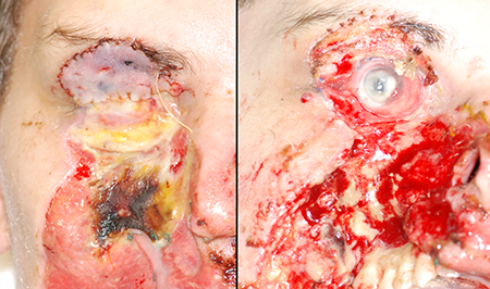
- Fourth-degree burns (deep burns): Similar severe damage to overlying tissue with underlying necrotic changes to muscle, bone and the deeper organs like the globe (Figure 2)

Figure 2. Patient 2 weeks after 4th-degree burn with loss of eyelids with developing eschar (left) status post conjunctival inverse advancement and postauricular skin graft done urgently for corneal melt. The graft failed and the patient ultimately underwent evisceration at the time of further eschar debridement several weeks later (right).
The pathology occurring at the epicenter of the injury is different than in the surrounding “zones”:
- Jackson’s burn injury model describes concentric zones starting with the zone of coagulation at the injury site, surrounded by the zone of stasis and the zone of hyperemia.
- A deep dermal burn can have surrounding partial thickness dermal burns and epidermal burns that both limit the healing of the epicenter and can also progress because of edema and infection.
Risk factors
Children and the elderly are most at risk given their relative immobility and dependence.
Similarly, the mentally disabled can be an at-risk population for burns.
Differential diagnosis
Infection such as preseptal cellulitis or necrotizing fasciitis
Patient management: treatment and follow-up
Natural history
Epidermal burns will heal without intervention in 5–7 days.
Superficial dermal burns often heal in 1–2 weeks with scarring or eyelid contracture a rare sequelae.
Deeper dermal burns require 2–3 weeks to heal and more often have scarring or contracture of the anterior lamella and loss of skin appendages such as hair follicles and sweat glands.
Full-thickness burns, owing to the damage to the deep regenerative dermis, heal from the wound edge inward. Contracture of the anterior lamella is near uniform and the consequence depends on the size of the burn.
Medical therapy
- Cleanse affected skin with moist gauze.
- Topical antibiotic can be applied to 1st- and 2nd-degree burns:
- Topical povidone-iodine ointment, silver sulfadiazine cream, silver nitrate solution and mupirocin ointment are commonly used around the face.
- Polysporin ophthalmic ointment can be used on the lids.
- Apply a lubricated dressing such as Xeroform gauze or alginate dressing.
- Trim burnt eyelashes with ointment covered scissors.
- Ocular-specific care:
- Sweep fornices with moist cotton tip applicators.
- For all patients with eyelid burns, give frequent preservative-free lubricating drops or ointment.
- If there is corneal injury, but no ulcer, add topical fluoroquinolone to cover pseudomonas.
- If there is corneal ulcer, add fortified topical antibiotics.
- Consider moisture chambers fashioned using ointment and smooth, occlusive dressing (e.g., Tegaderm) or simple plastic-wrap dressing.
- Boston scleral lens therapy has been investigated as a means of adding a layer of well-oxygenated fluid to cover the cornea and could be used if available.
- Application of a negative pressure dressing can be useful in deeply damaged facial burns, but care should be taken to avoid direct exposure to the ocular surface.
Surgery
Early surgical interventions are sometimes required for deeper burns:
- Suture tarsorrhaphy using bolsters to prevent cheese-wiring (IV tubing from a butterfly needle or pieces of a red rubber or Foley catheter are preferable to foam from a suture pack).
- A “draw-string” approach can be helpful to allow ophthalmic exam. It is fashioned by adding a second piece of bolstering material on the less-burned eyelid and leaving the tarsorrhaphy loose. The lids are kept closed by sliding the bolsters down and friction prevents them from sliding open.
- Suture tarsorrhaphy rarely holds in edematous or severely necrotic eyelids, and neither suture nor permanent tarsorrhaphies can prevent eyelid contracture from occurring, but are rather for protection of the globe.
- Lateral canthotomy and cantholysis should be performed in patients with orbital compartment syndrome with elevated intraocular pressure and/or afferent pupillary defects.
- In cases of loss of the eyelids or inability to perform tarsorrhaphy, temporizing measures to provide protection should be performed depending on the extent of the tissue damage, although these measures should not be considered permanent eyelid reconstructions.
- The ocular surface can be surgically covered with
- Any remaining viable conjunctiva. The peripheral bulbar or fornix conjunctiva can be mobilized and the upper and lower extent folded over and sewn to one another.
- Dual-layered amniotic membrane graft
- Split thickness dermal grafts
- Mobilized Tenon’s capsule
- The reconstructed surface can be covered by a temporary substrate such as
- Fresh or banked cadaveric skin
- Processed porcine skin (xenograft)
- Synthetic membrane (Biobrane)
- The reconstructed surface can be covered by a permanent substrate such as
- A full-thickness or partial-thickness skin graft
- Cultured epidermal cell sheets
- Synthetic membrane (Integra)
Intermediate stage surgical management is often required to alleviate lagophthalmos, release contractures, prevent malpositions, and treat infections.
- Grafting, although it might need to be repeated, should be entertained at this intermediate stage (1–3 weeks).
- Excision of necrotic eschar can be performed to prevent infection and aid in release of tissue, but it should be performed later than in other places of the body; 2–3 weeks is recommended.
- Contracture release and grafting has not conclusively been shown to be a benefit early (i.e., at 1 week) and should be delayed until corneal exposure forces the issue or when healing is felt to be complete.
- Consider depth and natural history discussion above.
- Unless contracture has resulted in complete loss of the anterior lamella, i.e., the brow skin is within 1 cm of the lash line, an incision should be made at the desired lid crease height and wide undermining should be performed to release all lid margin tension.
- A traction suture is useful for counter tension during dissection.
- In a 3rd-degree or milder 4th-degree upper-eyelid burn, dense scarring in the septum should be released to allow some mobility of the upper eyelid.
- A skin graft should be applied. Full-thickness grafts contract less owing to the increased dermal integrity. The graft should be oversized relative to the recipient site to account for contraction. Harvest locations are chosen based on available undamaged tissue of an adequate size in the following order of preference:
- Contralateral upper eyelid skin
- Preauricular skin
- Postauricular skin
- Supraclavicular skin
- Inner upper arm skin
- Split thickness grafting if needed for size
- The skin graft should be placed in a manner to decrease bleeding from limiting basal vascular ingrowth.
- Small drainage holes can be created using a #11 blade with or without a drain placement.
- The graft can be quilted centrally using interrupted tacking sutures or incorporated in the pass of a suspending Frost suture tarsorrhaphy.
- The graft can be glued using fibrin sealant such as Evicel or Tisseel glue.
- The lids should be immobilized on traction for 3–5 days.
- A pilot study has been published demonstrating some surgical success in 3rd degree burns of the upper eyelid using “very small autologous columnar skin grafts” harvested from a healthy site (Burns 2022:48:1671).
- Other malpositions that might need to be corrected include
- Horizontal laxity
- Canthal webs
- Composite eyebrow grafting
- Punctal stenosis
- Horizontal fissure shortening
Other management considerations
Corneal examination might dictate other ophthalmic interventions that need to be entertained. Corneal melting or chemical burn–related injuries might benefit from topical cyclosporine, topical doxycycline, or ascorbic acid.
Amniotic membrane transplantation for corneal epithelial defects is not necessary for mild burns. Its use in severe burns has been reviewed in a recent Cochrane review and not shown to have a clear benefit for corneal re-epithelialization.
Common treatment responses, follow-up strategies
In severe burns requiring early or intermediate grafting, multiple surgical procedures are common. Unless lagophthalmos is refractory and the globe is threatened, secondary grafting should be delayed 3–6 months.
Sun protection to burned skin of all types should be employed to limit the risk of scarring and secondary malignancy.
Burn- and surgical repair–related scars should be treated with massage, intralesional steroids such as triamcinolone 40 mg/mL, or intralesional antimetabolites such as 50 mg/ml 5-fluoruracil (off label).
Preventing and managing treatment complications
Failure to adequately protect the ocular surface can lead to corneal ulceration, endophthalmitis, and loss of the eye. As described above, various strategies to protect and cover the ocular surface should be employed.
Disease-related complications
- Corneal opacities from abrasions, ulcerations, limbal stem cell deficiency
- Corneal or scleral melting and endophthalmitis
- Conjunctival scarring can result in symblepharon, lid malposition, and motility deficits.
- Trichiasis with or without entropion
- Eyelid contracture and malposition such as entropion or ectropion with or without lagophthalmos
- Scarring of the skin with or without malposition is common in deep dermal and great burns.
- Burn scar malignancies such as basal cell and squamous cell carcinomas with difficult to quantify frequency
References and additional resources
- Malhotra R, Sheikh I, Dheansa B. The management of eyelid burns. Survey of ophthalmology 2009;54:356-71.
- Clare G, Suleman H, Bunce C, Dua H. Amniotic membrane transplantation for acute ocular burns. The Cochrane database of systematic reviews 2012;9:CD009379. http://www.ameriburn.org/resources_factsheet.php.
- Still JM, Jr., Law EJ, Belcher KE, Moses KC, Gleitsmann KY. Experience with burns of the eyes and lids in a regional burn unit. The Journal of burn care & rehabilitation 1995;16:248-52.
- Frye KE. Thermal Burns. In: J W, ed. Plastic Surgery Secrets Plus. 2nd ed. Philadelphia PA: Mosby Elsevier; 2010:643-65.
- Singh CN, Klein MB, Sullivan SR, et al. Orbital compartment syndrome in burn patients. Ophthalmic plastic and reconstructive surgery 2008;24:102-6.
- Hettiaratchy S, Dziewulski P. ABC of burns. Introduction. Bmj 2004;328:1366-8.
- Kalwerisky K, Davies B, Mihora L, Czyz CN, Foster JA, DeMartelaere S. Use of the Boston Ocular Surface Prosthesis in the management of severe periorbital thermal injuries: a case series of 10 patients. Ophthalmology 2012;119:516-21.
- Yao Z, Peng M, Liao J, Kong C, Fan S, Li C, Tang J, Chang D. Treatment of upper eyelid third-degree burns by dispersed implantation of very small autologous columnar skin grafts: A pilot study of a new method. Burns. 2022 Nov;48(7):1671-1679. doi: 10.1016/j.burns.2022.01.011. Epub 2022 Jan 22. PMID: 35221158.
