Silicone Intubation of Nasolacrimal Drainage System
Updated May 2024
Goals
- Dilate or maintain canaliculi
- Maintain surgical fistula following DCR
- Maintain patency of NLD following probing for congenital NLDO
- Treatment of acquired NLDO (controversial)
Indications and contraindications
Indications
Congenital dacryostenosis
- Relative lack of prospective comparative randomized studies
- Small case series claim variable success rates.
- Pediatric Eye Disease Investigator Group (PEDIG) studies (comprised of 200 pediatric ophthalmologists and pediatric optometrists in United States, Canada and the United Kingdom):
- Immediate in office vs. delayed facility probing: in a randomized trial (n = 57) no significant difference in success rate was found (AJO 2013;156:1045).
- Repeat probing: success rate (n = 20) was 56% (95% CI, 33%–76%) (J AAPOS 2009;13:306).
- Intubation vs. balloon catheter dilation: success was reported in 65 of 84 eyes (77%; 95% confidence interval, 65%–85%) in the balloon catheter dilation group and with 72 of 88 eyes (84% after adjustment for intereye correlation; 74%–91%) in the nasolacrimal intubation group. This difference was not statistically different (Arch Ophthalmol 2009;127:633).
- As a primary treatment in children 6 months to 4 years of age, overall success rate 91% (J AAPOS 2008;12:451).
- Primary treatment
- Usually reserved for children being treated that are over one year of age. No consensus on specific regarding at which age stenting should be used primarily (J AAPOS 2008;12:451).
- Not routinely recommended for children one year of age or less.
- Failed probing
- Success has been reported in up to 97% (Arch Ophthalmol 1998;116:387).
- Success rate is most consistently reported to lie between 80% and 85% (Am J Ophthalmol 1982;94:585, J Pediatr Ophthalmol Strabismus. 1980;17:389, J AAPOS 2004; 8:466, AJO 1994;118:781).
- In conjunction with balloon dacryoplasty (Arch Ophthalmol 2009;127:633).
- As primary procedure in selected patients, e.g., multifocal obstruction, canalicular stenosis noted on probing
- In conjunction with punctal reconstruction
- Technique variations have been described in the management of congenital obstruction
- Randomized comparative studies are lacking.
- Monocanalicular stenting (J AAPOS 2007; 11:183)
- Increased complication rate described (Ophthalmology 1998; 105:336)
- Double stenting (Ophthalmology 2008; 115:383)
Acquired nasolacrimal duct obstruction
- Partial/functional nasolacrimal duct obstruction (OPRS 2012;28:35).
- Acquired nasolacrimal duct obstruction
- Controversial with fairly low success rate, 25% to 50% in most series (Bull Soc Belge Ophthalmol 2008; 23:309, AJO 2007; 144:776, BJO 2006;90:435).
- In conjunction with dacryocystorhinostomy (DCR)
- Benefit has been challenged (Am J Rhinol. 2008;22:214, BJO 2009;93:1220; Ophthalmol. 2013; 120:2139-45).
-
Recent study shows advantage of intubation in endonasal DCR (Fayers T, Ophthalmology 2016)
In conjunction with trauma repair
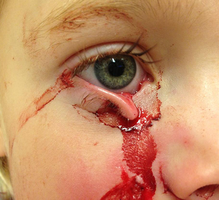
- Eyelid reconstruction following tumor resection (Figure 1)

Figure 1. Eyelid reconstruction following tumor resection.
Acquired canalicular obstruction
- In oncology patients treated with docetaxel (Taxotere) or 5-fluorouracil
- Patients with metastatic breast cancer treated with docetaxel, epiphora developed in 64% (n = 28) when administered weekly versus 39% (n = 28) when administered every 3 weeks (J Clin Oncol 2006; 24:3619).
- Some of these patients had mild stenosis that could be opened with office probe and irrigation.
- In half the patients with weekly docetaxel and 82% treated with docetaxel every 3 weeks, epiphora resolved with tobramycin-dexamethasone drops.
- The median cumulative dose of docetaxel at onset of epiphora was 420–496 mg.
- Referral to ophthalmologist should be with onset of epiphora to facilitate early intervention (Clin Breast Cancer 2005; 5:455)
- Begin with office dilation and Tobradex QID, with taper over 4 weeks if effective.
- For patients receiving docetaxel every 3 weeks, continue to monitor; might need repeat dilation and medical treatment; might need silicone intubation for significant progression.
- For patients receiving docetaxel weekly, consider early silicone intubation if stenosis significant and progressing.
- In 14 patients treated with weekly docetaxel, mean cumulative dose was 399 mg in patients with moderate canalicular stenosis and 501 mg with severe stenosis (Ann Oncol 2002;13:218)
- Docetaxel can be administered in cycles of 3 weekly infusions and 1 week off, and infusions are commonly accompanied by intravenous dexamethasone.
- The weekly dose ranges from 25–40 mg/m2
- Onset of epiphora ranged from 4 to 16 weeks (mean 7 weeks) after commencement of weekly docetaxel.
- Severe acquired canalicular stenosis secondary to chemotherapy may not improve with silicone intubation and may require Jones tube (Arch Ophthalmol 2001;119:1802).
Contraindications
- Complete canalicular/common canalicular obstruction without ability to restore patency: can proceed directly to conjunctivo-DCR
- Complete nasolacrimal duct obstruction is a relative contraindication with low success rate after silicone intubation; can proceed directly to DCR.
Preprocedure evaluation
- Patient history
- Periocular surgery including sinus and nasal and bony trauma
- Use of chemotherapy
- Placement of punctal plugs
- Clinical examination: cornea, conjunctiva, lid margin, punctum
- Complete lacrimal evaluation: dye disappearance test, canalicular probing/irrigation
Procedure alternatives
- Office probe and irrigation
- Dacryocystorhinostomy
- Conjunctivodacryocystorhinostomy (CDCR) with Jones tube placement
Surgical techniques
Intubation options
Monocanalicular stents
- Varieties
- With guide wire, retrieved from nasal cavity
- Without guide wire, pushed down canaliculi
- Benefit
- Retrieval in nose may not be required
- Can be performed without general anesthesia
- Easier removal, pulled out through puncta
- Disadvantage
- Smaller girth: single stent at level of common canaliculi and onward
- Seating in puncta may be less stable
Bicanalicular stents
- Varieties
- With long guide wire: retrieved with groove director or hemostat
- With olive tip: can be hooked in the nasal cavity
- Patent for threading of suture: Suture is pulled out nasal cavity dragging stent through lacrimal drainage duct.
- Pigtail (ring stent): looped through canaliculi system
- Variable thickness, with thin interpunctal segment
- Some brands attached to long olive tip probe
- Some brands with short guide wire that pushes stent into duct, then pulls out from above, avoiding the need to rescue the probe in the nose
- Benefit
- Secure: less likely be lost/fall out
- Can increased girth with two stents
- Disadvantage
- Can cause abnormal enlargement of puncta, i.e., cheese-wiring
- Can be challenging to remove
Anesthesia
General anesthesia
- Usually needed when stents are retrieved within the nasal cavity
Local anesthesia with/without sedation
Nasal cavity passage is too uncomfortable for most patients.
Monocanalicular stents (without the guide wire) can be placed under local anesthesia.
Nasal decongestion/vasoconstriction
- Useful for procedures involving entry into nasal cavity
Technique
Monocanalicular intubation
- Punctal dilation
- Variety
- No guide wire: simply push into distal canaliculi
- With guide wire: passed through lacrimal drainage system, same as with bicanalicular stents.
- Seating of punctal plug portion of monocanalicular stent in punctum, or suturing of free end of tube anterior to punctum
Bicanalicular intubation (ring intubation)
- Punctal dilation
- Introduction of pig-tail probe
- Careful passage of pig-tail probe through both canaliculi
- Placement of silicone tubing loop containing internal suture in lumen
- Tying of internal suture with rotation of tied ends of tubing into canaliculus
Bicanalicular intubation (nasolacrimal duct)
- Nasal decongestion/anesthesia
- Punctal dilation
- Passage of probe attached to tubing through canaliculus into lacrimal sac, and down nasolacrimal duct with retrieval of probe from inferior meatus (with/without endoscopy)
- Passage of second probe through other canaliculus in similar fashion
Tying/suturing of tubing ends in nasal vestibule
- +/- suturing of tubing knot to mucosa of lateral nasal wall
- A variation is to avoid knotting the stent, but rather join the 2 arms with a tight wrap-around absorbing suture, which is then tied to the nasal wall. When the suture absorbs, the stent will fall out or can be pulled out from above.
Bicanalicular intubation performed with DCR, etc.
- Passage of probes through canaliculi into DCR ostium
- Intranasal probe retrieval from ostium
- Tying/suturing/securing of tubes as above
Intubation with microendoscopic intracanalicular visualization
- Might decrease incidence of false passage, but equipment not widely available
Patient management: treatment and follow-up
Postoperative instructions
- Avoid manipulating stent.
- No eye-rubbing or nasal manipulation
- Possible use of eye shield at bedtime
- No/limited nose-blowing
- If stent fall out nose, push it back.
- If stent loops out palpebral fissure, proceed to clinic.
Medications prescribed
- Systemic antibiotics (variable use among surgeons)
- Topical antibiotics (variable use among surgeons)
- Topical steroids (variable use among surgeons)
Other management considerations
- Evaluate patient periodically to assess for stretching (cheese wiring) of puncta/canaliculi or interpunctal segment that is too tight.
- Office exam regarding presence of below complications
Stent removal
Timing
- Comparative studies needed
- Usually 3 to 4 months
- Practice patterns highly variable
Location
- Office
- Operating room (OR)/anesthesia might be needed for children
Direction
- Transnasal: identify, cut, and pull out nose.
- Transcanalicular removal: If a small knot was used or an absorbing suture with no knot in the stent, it can be pulled out canaliculi and puncta.
Preventing and managing treatment complications
Complications and preventive measures
- Recurrent/progressive dacryostenosis
- Cheese-wiring through puncta/canaliculi
- Appropriate tension on tubing loop in medial commissure: Tube should be visible between the upper and lower lid margins.
- Monitor and remove is cheese-wiring is identified
- Infection
- False passage creation
- Stent is passed outside normal lacrimal drainage path.
- Might occur at level of obstruction
- High risk with canaliculi obstruction
- Stent might exit wrong location in nose.
- Medial meatus if passed though nasal wall
- Submucosal if passed through lacrimal sac mucosa
- Epistaxis
- Allergy to stent
Management of complications
- Recurrent/progressive dacryostenosis: appropriate surgery
- Cheese-wiring through puncta/canaliculi
- Stent removal
- Canalicular repair
- Possible stent replacement under less tension
- Infection
- Stent removal
- +/- antibiotic treatment
- False passage creation
- Stent removal
- Possible antibiotic treatment/additional surgery
- Stent dislodged
- If the nasal ends are showing, they can be trimmed.
- If the stent dislodges and is hanging out of the eye, it can sometimes be repositioned with a lacrimal probe from above. (Ophthalmic Surg. 1985;16(5):307-8) Otherwise, it might be possible to identify the end beneath the inferior turbinate and pull the stent back in position.
- If the tube falls out completely, reassure the patient and let them know that often, when the tube falls out, it is because the system has opened and the procedure has been successful.
References and additional resources
- AAO, Basic and Clinical Sciences Course. Section 7: Orbit, Eyelids, and Lacrimal System, 2013-2014.
- AAO, Surgery of the Eyelid, Orbit & Lacrimal system, Vol. 3, 1995, p. 275.
- AAO, Focal Points: Evaluation and Surgery of the Lacrimal Drainage System in Adults, Module #12, 1995, p. 12-13.
- Ahmadi MA, Esmaeli B. Surgical treatment of canalicular stenosis in patients receiving docetaxel weekly. Arch Ophthalmol. 2001;119:1802-4.
- Bleyen I, Paridaens AD. Bicanalicular silicone intubation in acquired partial nasolacrimal duct obstruction. Bull Soc Belge Ophtalmol. 2008;(309-310):23-6.
- Bleyen I, van den Bosch WA, Bockholts D, Mulder P, Paridaens D. Silicone intubation with or without balloon dacryocystoplasty in acquired partial nasolacrimal duct obstruction. Am J Ophthalmol. 2007;144:776-780.
- Chong KK, Lai FH, Ho M, Luk A, Wong BW, Young A. Randomized trial on silicone intubation in endoscopic mechanical dacryocystorhinostomy (SEND) for primary nasolacrimal duct obstruction. Ophthalmol. 2013; 120:2139-45.
- Connell PP, Fulcher TP, Chacko E, O’ Connor MJ, Moriarty P. Long term follow up of nasolacrimal intubation in adults. Br J Ophthalmol. 2006;90:435-6.
- Demirci H, Elner VM. Double silicone tube intubation for the management of partial lacrimal system obstruction. Ophthalmology 2008;115:383-5.
- Dortzbach RK, France TD, Kushner BJ, Gonnering RS. Silicone intubation for obstruction of the nasolacrimal duct in children. Am J Ophthalmol 1982;94:585-90.
- Durso F, Hand SI Jr, Ellis FD, Helveston EM. Silicone intubation in children with nasolacrimal obstruction. J Pediatr Ophthalmol Strabismus. 1980;17:389-93.
- Engel JM, Hichie-Schmidt C, Khammar A, Ostfeld BM, Vyas A, Ticho BH. Monocanalicular silastic intubation for the initial correction of congenital nasolacrimal duct obstruction. J AAPOS 2007;11:183-6.
- Esmaeli B, Amin S, Valero V, Adinin R, Arbuckle R, Banay R, Do KA, Rivera E. Prospective study of incidence and severity of epiphora and canalicular stenosis in patients with metastatic breast cancer receiving docetaxel. J Clin Oncol. 2006;24:3619-22.
- Esmaeli B, Golio D, Lubecki L, Ajani J. Canalicular and nasolacrimal duct blockage: an ocular side effect associated with the antineoplastic drug S-1. Am J Ophthalmol. 2005;140:325-7.
- Esmaeli B, Hortobagyi G, Esteva F, Valero V, Ahmadi MA, Booser D, Ibrahim N, Delpassand E, Arbuckle R. Canalicular stenosis secondary to weekly docetaxel: a potentially preventable side effect. Ann Oncol. 2002;13:218-21.
- Esmaeli B. Management of excessive tearing as a side effect of docetaxel. Clin Breast Cancer. 2005 Feb;5(6):455-7.
- Fayers T, Dolman PJ. Bicanalicular silicone stents in endonasal dacryocystorhinostomy: results of a randomized clinical trial. Ophthalmology 2016;123:2255-9.
- Gonnering RS. Gentle, technically simple repositioning of displaced lacrimal tubing. Ophthalmic Surg. 1985 May;16:307-8.
- Heirbaut AM1, Colla B, Missotten L. Silicone intubation for congenital obstruction of nasolacrimal ducts. Bull Soc Belge Ophtalmol 1990;238:87-93.
- Kaufman LM, Guay-Bhatia LA. Monocanalicular intubation with Monoka tubes for the treatment of congenital nasolacrimal duct obstruction. Ophthalmology 1998;105:336-41.
- Lee KA, Chandler DL, Repka MX, Melia M, Beck RW, Summers CG, Frick KD, Foster NC, Kraker RT, Atkinson S; PEDIG. A comparison of treatment approaches for bilateral congenital nasolacrimal duct obstruction. Am J Ophthalmol 2013;156:1045-50.
- Lim CS, Martin F, Beckenham T, Cumming RG. Nasolacrimal duct obstruction in children: outcome of intubation. J AAPOS. 2004 Oct;8(5):466-72.
- Moscato EE, Dolmetsch AM, Silkiss RZ, Seiff SR. Silicone intubation for the treatment of epiphora in adults with presumed functional nasolacrimal duct obstruction. Ophthal Plast Reconstr Surg. 2012;28:35-9.
- Pe MR, Langford JD, Linberg JV, Schwartz TL, Sondhi. Ritleng intubation system for treatment of congenital nasolacrimal duct obstruction. Arch Ophthalmol 1998;116:387-91
- Pediatric Eye Disease Investigator Group, Repka MX, Melia BM, Beck RW, Chandler DL, Fishman DR, Goldblum TA, Holmes JM, Perla BD, Quinn GE, Silbert DI, Wallace DK. Primary treatment of nasolacrimal duct obstruction with balloon catheter dilation in children younger than 4 years of age. J AAPOS 2008;12:451-5.
- Ratliff CD, Meyer DR. Silicone intubation without intranasal fixation for treatment of congenital nasolacrimal duct obstruction. Am J Ophthalmol. 1994;118:781-5.
- Repka MX, Chandler DL, Bremer DL, Collins ML, Lee DH. Repeat probing for treatment of persistent nasolacrimal duct obstruction. J AAPOS 2009;13:306-7.
- Repka MX, Chandler DL, Holmes JM, Hoover DL, Morse CL, Schloff S, Silbert DI, Tien DR; Pediatric Eye Disease Investigator Group. Balloon catheter dilation and nasolacrimal duct intubation for treatment of nasolacrimal duct obstruction after failed probing. Arch Ophthalmol 2009;127:633-9.
- Saiju R, Morse LJ, Weinberg D, Shrestha MK, Ruit S. Prospective randomised comparison of external dacryocystorhinostomy with and without silicone intubation. Br J Ophthalmol. 2009;93:1220-2.
- Smirnov G, Tuomilehto H, Teräsvirta M, Nuutinen J, Seppä J. Silicone tubing is not necessary after primary endoscopic dacryocystorhinostomy: a prospective randomized study. Am J Rhinol. 2008;22:214-7.
