Sinus Mucocele
Updated May 2024
Establishing the diagnosis
Etiology
- Mucus-filled cystic structures with pseudostratified ciliated columnar (respiratory) epithelium
- Results from obstruction of the sinus excretory ducts
Epidemiology
- 70% involve the frontal and ethmoid sinuses; less commonly the maxillary (5%–10%) and sphenoid sinuses (2%).
- 28% present with orbital manifestations, which is the leading presentation after headache.
- Up to 70% of patients with mucocele initially present with ophthalmic signs and symptoms.
- Mucoceles represent 4% of all orbital mass lesions.
- Thinning of the bony wall over time might allow for extension into the orbit and other surrounding structure.
- Most cases involve adults, but young children with cystic fibrosis are also prone to develop mucoceles.
- Might be the presenting sign of cystic fibrosis
History
- Initial onset and duration of symptoms
- Change in vision
- Diplopia
- Hypesthesia
- Pain
- Epiphora
- Nasal congestion or discharge
- Headache
Clinical features
Mass effect
- Often a palpable mass distorting the periocular structures.
- If anterior bony wall is present, the mass will be firm.
- If anterior bony resorption has occurred, the mass will be softer.
- The mass is usually not tender, and skin is intact.
- Depending on the sinus, can cause a superior, medial, or inferior mass effect.
- If mucocele communicates with intracranial vault, the mass can be pulsatile.
Periorbital edema
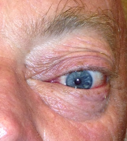
Proptosis and globe displacement common with orbital invasion (Figure 1)
Direction of globe displacement depends on location of mucocele.
Orbital compression—less common
- Diplopia (13%)
- Motility restriction (4.6%)
- Secondary to mass effect on extraocular muscles or oculomotor nerve palsy from compression on the cranial nerves
- Optic neuropathy (4%–18%)
- More common with sphenoid sinus and posterior ethmoidal (Onodi cell) mucoceles due to close proximity to the orbital apex
- Much less common with frontoethmoidal mucoceles, although possible with large mucoceles that have been neglected over time
- Many of these are reversible with decompression of the orbital apex by removal of the mucocele.
- Poor outcome more likely for cases of long-standing neuropathy and cases of mucopyocele

Figure 1. External photograph showing left proptosis and hyperglobus.
Testing
External exam looking for changes in facial bony architecture
Ophthalmic exam focusing on
- Visual acuity
- Color vision
- Pupils, including afferent pupillary defect
- Motility
- Exophthalmometry
- Globe dystopia
- The direction of dystopia depends on the sinus affected.
- The frontal mucocele will cause inferior globe dystopia.
- DFE with attention to the optic nerve
- Displacement of the globe might also result in chorioretinal folds.
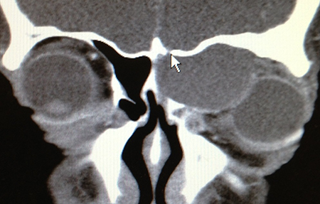
CT scan (Figure 2)
- Capsulated mass with homogeneous density, isodense with cerebral tissue (similar tissue density)
- Affected sinus is completely opacified; peripheral calcification is sometimes seen.
- Peripheral enhancement after injection of contrast
- Can identify bony defects and extension into adjacent structures
- Overlying sinusitis often difficult to see on imaging.
- If sinusitis extends to adjacent sinuses, will see opacification in those sinuses.
MRI
- Variable appearance, depending on the state of hydration of mucus-filled cysts
- Can present hyperintense in T1 because of high protein concentration in the mucus
- Can present hyperintense in T2 because of large quantity of water
- Can present as hypointense in T1 and absent in T2 because of dry mucus

Figure 2. Coronal CT image of the same patient showing a left ethmoidal mucocele with invasion into the superomedial orbit. The arrow points to a small area of bone erosion superiorly with communication into the cranial fossa.
Risk factors
- Previous trauma
- Previous sinus surgery
- Sinus inflammation/allergies
- Neoplasm
- Iatrogenic
Differential diagnosis
- Neoplasm
- Infection
- Encephalocele
- Trauma
Patient management: treatment and follow-up
Natural history
- Slow growth over time as mucus production continues into the cyst
- Can extend into surrounding structures, including the nasopharynx, cranium, and orbit
- Secretion of prostaglandins and collagenases, which allow for bone resorption
- Although not a life-threatening condition, left untreated, mucocele can cause
- Disfiguring proptosis or enophthalmos
- Soft tissue distortion in the periocular area
- Epiphora
- Lower-lid malposition
- Ptosis
- Motility restriction
- Optic nerve compression
Medical therapy
- Intranasal steroids
- Antibiotics
Surgery
Surgical treatment includes evacuation of the mucocele and re-establishment of drainage of the affected sinus or obliteration of the sinus by mucosal stripping and packing with bone, fat, or fascia.
Surgical approach can be separated into external or endoscopic.
- These have comparable rates in terms of complications and recurrence.
- The choice depends on the comfort level of the surgeon and the extent of the mucosal disease.
- Although endoscopic approaches are most commonly used today, external approaches can be used
- When previous endoscopic approaches have failed
- In cases with difficult anatomy, which hampers the endoscopic approach
- When the advanced instrumentation required for endoscopy is not available
External approaches
Frontal sinus mucocele:
- Can be approached by a coronal incision or Lynch incision
- A frontal bone flap overlying the mucocele is then created, and the mucocele is removed.
- When completed, the bone flap is then resecured into place with rigid fixation.
- The sinus mucosa can then be removed, and the frontal sinus duct closed with abdominal fat or fascia, commonly from the frontalis muscle.
- Appropriate when the posterior table of the frontal sinus is intact
- This should be left intact to prevent CSF leak.
- Advantage of the external approach is wide visualization of the sinus.
- Can also be addressed by the Lothrop procedure, which involves removal of the frontal sinus floor, intersinus septum, and superior nasal septum
- This procedure is now performed almost exclusively through an endoscopic approach (modified Lothrop or Draf III procedure).
- Creates a new outflow tract for the frontal sinus rather than obliteration of the sinus
- Alternatively, the frontal sinus can be cranialized through a craniotomy by removing the posterior wall of the frontal sinus.
- This is preferred when there is posterior extension of the mucosa into the cranial vault with thinning or absence of the posterior sinus wall.
- The mucosa is removed, and the brain and dura are allowed to fill the space.
- The anterior sinus wall is left intact.
- Should be done in conjunction with a neurosurgeon
- Allows for preservation of the normal appearance of forehead contour
- Complications include CSF leak, intracranial infection, and mucosal regrowth.
Ethmoid sinus mucocele can be approached from a Lynch or transcaruncular incision.
Maxillary sinus mucocele can be approached from a Caldwel Luc or lateral rhinotomy.
Sphenoid sinus mucocele can be approached from a craniotomy.
- Should be done in conjunction with a neurosurgeon
Endoscopic approach
All of the above sinuses can be treated through an endonasal endoscopic procedure.
- Once the mucosa is removed, the osteum of the sinus is opened widely to allow for egress of the mucosal secretions.
- These techniques require specialized instruments, cameras, and advanced training.
- Unless endoscopic comfort level is high, these should be done in conjunction with an endoscopic trained ENT surgeon.
Advantages:
- Reduced postoperative morbidity
- Lack of external incision
- Preserved forehead sensation.
This approach is safe and effective even with orbital extension of the mucocele.
Other management considerations
Is an orbital approach always necessary concurrently with sinus surgery for mucoceles that invade the orbit?
- Not always; most of the orbital signs are due to mass effect into the orbit by the mucocele.
- Simply removing the mucocele often leads to resolution of the orbital signs.
- Cases in which an orbital approach would be useful include
- Drainage of an intraorbital abscess associated with a mucopyocele
- Cases in which the sinus surgeon does not have a high comfort level evacuating a mucocele that abuts important orbital structures (optic nerve, globe)
- In these cases, approach from the orbital side can allow for protection of important structures and help the sinus surgeon with intraoperative orientation.
Is orbital reconstruction necessary at the time of mucocele removal?
- Not always; Shah et al. demonstrated in select cases, removal of the mucocele alone is curative of the orbital signs.
- Even in cases with bone destruction, the body is capable of remodeling the bony defect.
- It should be noted that none of the patients in the above study had evidence of optic neuropathy or infection on presentation.
- The decision about whether and when to proceed with orbital reconstruction should be individualized.
- It is reasonable in the majority of cases to allow for a period of observation after mucocele removal.
- Orbital reconstruction can then be planned secondarily for patients with persistent globe displacement or diplopia from their orbital wall defect.
- Primary reconstruction should be considered in patients with large orbital wall defects who are not good candidates for multiple surgical procedures due, for instance, to medical comorbidities or living far away.
Common treatment responses, follow-up strategies
After surgical excision, mucoceles recur in 10%–26% of cases.
Monitor with periodic exams and imaging.
Prognosis for both life and vision is typically excellent with appropriate management.
Long-standing cases with compression on the optic nerve can result in optic atrophy and permanent vision loss.
Preventing and managing treatment complications
- Recurrence
- Complications of external approach include
- Scar formation
- Numbness
- Longer surgical time
- Infection
- If fat is harvested, complications include
- Donor site morbidity
- Difficulty with postoperative monitoring due to the appearance of fat on MRI, which can mask early recurrence
- Endoscopic approach carries a small risk of
- Hemorrhage
- Intracranial injury
- Damage to the orbital structures
Disease-related complications
- Mucopyocele, which can lead to orbital cellulitis
- Permanent vision loss from prolonged optic nerve compression (rare)
- Permanent motility restriction from damage to extraocular muscles or cranial nerves (rare)
- When neglected, can cause
- Disfiguring proptosis or enophthalmos
- Epiphora
- Lower-lid malposition
- Ptosis
Historical perspective
Frontal mucocele was first described by Dezeimeris in 1725.
In 1818, Litersangeback described signs and symptoms of mucoceles, which he called “hydatidic cyst.”
The term “mucocele” was first used by Rollet in 1896 to describe a lesion in the superior orbit.
Although external approaches used to be the mainstay of treatment, Kennedy’s landmark paper on treatment of mucoceles by endoscopic marsupialization in 1989 created a shift in the management of these cases.
With advances in computer guided imaging, cameras, and instrumentation, most authors in the literature now favor an endoscopic approach.
References and additional resources
- Berthon E. Essai sur les abces et hydropsies des sinus frontaux. Thesis, Paris, 1880.
- Courson AM, Stankiewicz JA, Lal D. Contemporary management of frontal sinus mucoceles: a meta-analysis. Laryngoscope. 2014 Feb;124(2):378-86.
- Devars du Mayne M, Moya-Plana A, Malinvaud D, et al. Sinus mucocele: natural history and long-term recurrence rate. Eur Ann Otorhinolaryngol Head Neck Dis. 2012;129(3):125-30.
- Draf W. Endonasal micro-endoscopic frontal sinus surgery: The Fulda concept. Operative techniques in Otolaryngology-Head and Neck Surgery. 1991;4:234-240.
- Guttenplan MD, Wetmore RF. Paranasal sinus mucocele in cystic fibrosis. Clin Pediatr (Phila). 1989;28(9):429-30.
- Iliff CE. Mucoceles in the orbit. Arch Ophthalmol. 1973 May;89(5):392-5.
- Kennedy DW, Josephson JS, Zinreich SJ, et al. Endoscopic sinus surgery for mucoceles: a viable alternative. Laryngoscope 1989;99:885–895.
- Kim YS, Kim K, Lee JG, et al. Paranasal sinus mucoceles with ophthalmologic manifestations: a 17-year review of 96 cases. Am J Rhinol Allergy. 2011 Jul-Aug;25(4):272-5.
- Langeback CJM. Neue Bibliothek fur die Chirurgie und Ophthalmologie. Hahn, Hannover, 1818.
- Leventer DB, Linberg JV, Ellis B. Frontoethmoidal mucoceles causing bilateral chorioretinal folds. Arch Ophthalmol. 2001 Jun;119(6):922-3.
- Lloyd G, Lund VJ, Savy L, Howard D. Radiology on focus. Optimum imaging for mucoceles. J Laryngol Otol 2000;114:233–236.
- Loo JL, Looi AL, Seah LL. Visual outcomes in patients with paranasal mucoceles. Ophthal Plast Reconstr Surg. 2009 Mar-Apr;25(2):126-9.
- Malhotra R, Wormald PJ, Selva D. Bilateral dynamic proptosis due to frontoethmoidal sinus mucocele. Ophthal Plast Reconstr Surg. 2003 Mar;19(2):156-7.
- McLeod J, Lux P. Cicatricial ectropion as a result of mucocele of the frontal sinus. Arch Ophthalmol. 1936;15(6):994-997.
- Moriyama H, Nakajima T, Honda Y. Studies on mucocoeles of the ethmoid and sphenoid sinuses: Analysis of 47 cases. J Laryngol Otol. 1992;106:23–27.
- Ormerod LD, Weber AL, Rauch SD, Feldon SE. Ophthalmic manifestations of maxillary sinus mucoceles. Ophthalmology. 1987 Aug;94(8):1013-9.
- Qureishi A, Lennox P, Bottrill I. Bilateral maxillary mucoceles: an unusual presentation of cystic fibrosis. J Laryngol Otol. 2012;126(3):319-21.
- Reinecke RD, Montgomery WW. Oculomotor nerve palsy associated with mucocele of the sphenoid sinus. Arch Ophthalmol. 1964 Jan;71:50-1.
- Rinna C, Cassoni A, Ungari C, et al. Fronto-orbital mucoceles: our experience. J Craniofac Surg. 2004 Sep;15(5):885-9.
- Rollet M. Mucocele de l’angle superointerne des orbites. Lyon Med 81:571–576,18, 1896.
- Sautter NB, Citardi MJ, Perry J, Batra PS. Paranasal sinus mucoceles with skull-base and/or orbital erosion: is the endoscopic approach sufficient? Otolaryngol Head Neck Surg. 2008 Oct;139(4):570-4.
- Shah A, Meyer DR, Parnes S. Management of frontoethmoidal mucoceles with orbital extension: is primary orbital reconstruction necessary? Ophthal Plast Reconstr Surg. 2007 Jul-Aug;23(4):267-71.
- Silverman JB, Gray ST, Busaba NY. Role of osteoplastic frontal sinus obliteration in the era of endoscopic sinus surgery. Int J Otolaryngol 2012;2012:501896.
- Soyka MB, Annen A, Holzmann D. Where endoscopy fails: indications and experience with the frontal sinus fat obliteration. Rhinology 2009;47:136–140.
- Valvassori GE, Putterman AM. Ophthalmological and radiological findings in sphenoidal mucoceles. Arch Ophthalmol. 1973 Dec;90(6):456-9.
- Weber R, Draf W, Keerl R, et al. Osteoplastic frontal sinus surgery with fat obliteration: technique and long-term results using magnetic resonance imaging in 82 operations. Laryngoscope 2000;110:1037–1044.
