Surgical Approaches to the Orbit
Updated July 2024
Indications and contraindications to orbitotomy
Indications
- Biopsy of orbital lesion suspected to be malignant
- Biopsy of orbital process in which tissue is needed for diagnosis
- Removal of orbital tumor/lesion causing symptoms or threatening vision
- Repair of orbital fracture
- Thyroid orbitopathy or other congestive disorder requiring decompression
- Removal of high-risk orbital foreign body
- Drainage or debridement of orbital abscess/infection
- Correction of proptosis or enophthalmos
- Reconstruction of orbital deformity
- Idiopathic intracranial hypertension or other condition requiring optic nerve sheath fenestration
- Exploration for lost rectus muscle
Contraindications
- Relative
- Medical risks for anesthesia
- Anticoagulation – Consider relative risks/benefits of stopping anticoagulation
- Presence of systemic infection
- Absolute
- Inability to tolerate anesthesia
Pre-procedure evaluation
- Complete medical/ophthalmic history with special attention to:
- Malignancy
- Autoimmune disease
- Prior ocular/orbital surgery or trauma
- Anticoagulation
- Risk factors for anesthesia complications
- Clinical examination
- Visual acuity
- Exophthalmometry
- Globe malposition
- Extraocular motility
- Optic nerve function
- Afferent pupillary defect
- Color vision
- Visual field
- Optic disc appearance
- Palpable masses
- Inflammatory signs
- Preoperative assessment
- Orbital imaging
- Computed tomography (CT) scan
- Magnetic resonance imaging (MRI)
- Angiography/venography
- Ultrasound
- Use of antibiotics, perioperative steroids
- Preop antibiotics
- One time dose intravenous Decadron 8 – 10 mg
- Anesthesia (most orbitotomies performed under general)
- Consider relative risks/benefits of stopping anticoagulation
- Equipment considerations
- Cryoprobe: useful for extraction of encapsulated tumors, particularly intraconal (Rosen, Acta Ophthalmol 2010)
- Endoscope: necessary for transnasal approach, helpful for visualization of apex
- Intraoperative navigation
- Intraoperative imaging (X-ray, ultrasound, fluoroscopy, CT, MRI)
- Implants
- Procedure-specific needs
- Fracture fixation hardware
- Osteotomes/ronguers
- Drills
- Ultrasonic bone aspirator
- Orbital imaging
Alternatives to orbitotomy
- Observation
- Medical treatment
- Steroids – thyroid orbitopathy, idiopathic orbital inflammation, compressive opticneuropathy
- Radiation – malignant tumors, thyroid orbitopathy, orbital inflammation
- Chemotherapy – malignant tumors
- Immunosuppressants – inflammatory disorders
- Minimally invasive procedures
- Sclerotherapy – lymphatic malformations (Hill, OPRS 2012)
- Embolization – vascular anomalies
- Fine needle aspiration – tumor biopsy
Surgical techniques
Transconjunctival approaches (Figure 1)
- Inferior fornix incision (McCord, Ophthalmic Surg 1979)
- Access to inferior/medial/lateral orbit for:
- Orbital floor fracture repair
- Repair of zygomaticomaxillary buttress fracture
- Exploration/biopsy/removal of inferior orbital lesion/foreign body
- Placement of enophthalmos wedge
- Lower lid blepharoplasty with fat removal
- Technique:
- Horizontal incision made below inferior border of tarsus
- Lateral canthotomy/inferior cantholysis can be performed to improve exposure (“swinging eyelid” approach)
- Lower lid retractors divided
- Dissection can be carried in preseptal or postseptal (Bernardini, OPRS 2017) plane
- For subperiosteal work, periosteal incision made along or just behind inferior rim
- Avoid damage to inferior oblique origin
- Conjunctiva closed with absorbable sutures
- Access to inferior/medial/lateral orbit for:
- Transcaruncular incision (Shorr, Ophthalmology 2000)
- Access to medial/inferior/superior orbit for:
- Medial orbital decompression
- Medial orbital wall fracture repair
- Drainage of subperiosteal abscess
- Exploration/biopsy/removal of medial orbital lesion/foreign body
- Exploration for lost medial rectus muscle (Goldberg, Am J Ophthalmol 2001)
- Technique:
- Vertical incision made through, lateral, or medial to caruncle
- Blunt dissection carried to posterior lacrimal crest
- For subperiosteal work, periosteal incision made behind posterior lacrimal crest
- Anterior and posterior ethmoidal vessels can be cauterized/divided if needed
- Incision closed with buried absorbable suture or allowed to heal without sutures
- Access to medial/inferior/superior orbit for:
- Peritomy incision
- Access to subtenon’s space, rectus muscles, intraconal space for:
- Strabismus surgery
- Enucleation
- Globe exploration
- Optic nerve sheath fenestration (Galbraith, Am J Ophthalmol 1973)
- Exploration/biopsy/removal of intraconal lesion/foreign body
- Technique:
- Incision made through conjunctiva/anterior Tenon’s capsule at limbus
- Subtenon’s dissection in quadrant(s) of interest
- Rectus muscle(s) can be tagged with vicryl sutures and detached from globe to improve exposure
- Blunt dissection carried into muscle cone
- Rectus muscle(s) reattached to globe at case conclusion
- Conjunctiva closed with absorbable sutures
- Access to subtenon’s space, rectus muscles, intraconal space for:
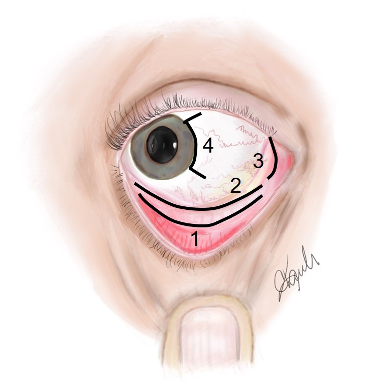
Figure 1. Illustration of various transconjunctival orbitotomy incisions, right eye. 1. Inferior transconjunctival, preseptal approach; 2. Inferior transconjunctival (fornix), postseptal approach; 3. Transcaruncular; 4. Nasal peritomy.
Transcutaneous approaches (Figure 2)
- Upper lid crease incision
- Access to superior orbit/rim, brow, supraorbital nerve, trochlea for:
- Exploration/biopsy/removal of superior orbital lesion/foreign body
- Drainage of subperiosteal abscess
- Repair of orbital roof fracture
- Removal of orbital fat
- Technique:
- Incision through anterior lamella at level of lid crease
- Orbital septum opened at desired location
- For subperiosteal work, preseptal dissection carried to superior orbital rim and periosteum opened over rim
- Supraorbital bundle should be avoided
- Closure as for blepharoplasty
- Access to superior orbit/rim, brow, supraorbital nerve, trochlea for:
- Lateral upper lid crease incision (Harris, OPRS 1999)
- Access to lateral/superior orbit, intraconal space for:
- Lateral orbital decompression
- Repair of lateral rim/zygomaticomaxillary complex fracture
- Exploration/biopsy/removal of lateral/intraconal orbital lesion/foreign body
- Lacrimal gland biopsy/removal/resuspension
- Removal of greater sphenoid wing for transorbital access to cavernous sinus/middle cranial fossa (Chabot, Neurosurgery 2017)
- Technique:
- Incision made through lateral upper lid crease
- For subperiosteal work, periosteum opened over lateral orbital rim
- Zygomaticofacial and zygomaticotemporal bundles can be cauterized/divided as needed
- Temporalis muscle can be dissected from zygoma/sphenoid if access to temporal surface of lateral wall needed
- Lateral orbital rim can be removed to improve exposure, refixated at end of case
- Skin incision closed with sutures
- Access to lateral/superior orbit, intraconal space for:
- Medial upper lid crease incision (Pelton, OPRS 2001)
- Access to superior/medial orbit, intraconal space for:
- Optic nerve sheath fenestration
- Exploration/biopsy/removal of superomedial or intraconal lesion/foreign body
- Cannulation of superior ophthalmic vein for cavernous sinus embolization (Miller, J Neurosurg 1995)
- Technique:
- 10mm incision made through medial upper lid crease
- Orbital septum opened, blunt dissection carried between superior oblique tendon and medial horn of levator aponeurosis
- Approach avoids ciliary ganglion, which sits on lateral aspect of optic nerve
- Skin incision closed with sutures
- Access to superior/medial orbit, intraconal space for:
- Upper lid split (Kersten, OPRS 1999)
- Access to superior orbit, intraconal space for exploration/biopsy/removal of intraconal lesion/foreign body
- Technique:
- Vertical full-thickness incision made through medial upper lid, extended superiorly through upper lid retractors/conjunctiva
- Dissection carried past superior oblique tendon through intramuscular septum between superior and medial rectus muscles
- Closed with meticulous repair of full-thickness lid laceration
- Lateral canthotomy/inferior cantholysis
- Access to lateral/inferior orbit for:
- Lateral tarsal strip procedure
- Emergent treatment of orbital compartment syndrome
- Exploration/biopsy/removal of lateral orbital lesion/foreign body
- Improvement of exposure during inferior transconjunctival orbitotomy (“swinging eyelid”)
- Technique:
- Horizontal incision from lateral commissure to lateral orbital rim (canthotomy)
- Vertical incision through inferior limb of lateral canthal tendon (cantholysis)
- In routine cases, lateral canthal tendon is resuspended from periosteum over lateral orbital tubercle with buried absorbable suture
- Lateral canthal angle reformed with buried absorbable suture
- Access to lateral/inferior orbit for:
- Lateral canthotomy (Hamed-Azzam, Eye 2017)
- Access to lateral orbit/intraconal space for exploration/biopsy/removal of lateral/intraconal lesion/foreign body
- Technique:
- Horizontal incision created from lateral commissure to lateral orbital rim
- Traction sutures placed through both limbs of lateral canthal tendon
- Blunt dissection carried in to lateral orbit/intraconal space
- LCT/skin reapproximated at conclusion of case
- Lateral canthus incision (modified Berke)
- Access to lateral orbit for decompression or exploration/biopsy/removal of lateral lesion/foreign body
- Technique:
- Horizontal incision from lateral orbital rim to temporalis fossa, sparing lateral canthal tendon
- Lateral orbitotomy proceeds in similar fashion to lateral lid crease incision
- Uncommonly performed
- Other lateral orbitotomy incisions (historical, rarely performed)
- Krönlein – horseshoe shaped incision over lateral orbital rim
- Stallard-Wright – s-shaped incision from lateral brow to lateral canthus/temporalis fossa
- Berke – lateral canthotomy extending from lateral commissure horizontally over lateral canthus/temporalis fossa
- Subciliary incision
- Access to inferior rim/orbit for:
- Orbital floor fracture repair
- Lower lid blepharoplasty with fat removal/repositioning
- Technique:
- Horizontal incision made across lower lid just beneath lash line
- Incision can be extended inferolaterally through rhytid to improve exposure or facilitate skin removal for blepharoplasty
- Anterior lamella dissected from septum
- For subperiosteal work, periosteum opened over inferior orbital rim
- Incision closed as for blepharoplasty
- Access to inferior rim/orbit for:
- Medial canthal tendon (Nunery) incision (Timoney, OPRS 2012)
- Access to lacrimal sac fossa and medial orbit for:
- Medial orbital wall fracture repair
- Medial orbital decompression
- Naso-orbital-ethmoid fracture repair
- Exploration/biopsy/removal of medial orbital lesion/foreign body
- Technique:
- 15-20mm incision created over anterior crus of medial canthal tendon
- MCT disinserted from anterior lacrimal crest
- Lacrimal sac dissected from lacrimal sac fossa
- Dissection carried past posterior lacrimal crest into medial orbit
- Incision closed with skin sutures
- Access to lacrimal sac fossa and medial orbit for:
- Lynch incision (Lynch, Laryngoscope 1921)
- Access to medial orbit for:
- Repair of naso-orbital-ethmoid fractures
- Repair of medial orbital wall fractures
- Drainage of medial subperiosteal abscess
- Technique:
- Curvilinear incision made over medial orbital rim
- Z-plasty modification can improve wound cosmesis (Esclamado, Laryngoscope 1989)
- Medial canthal tendon disinserted
- Subperiosteal dissection carried over lacrimal sac fossa and medial orbit
- Skin incision closed with sutures
- Rarely utilized by oculofacial surgeons
- Access to medial orbit for:
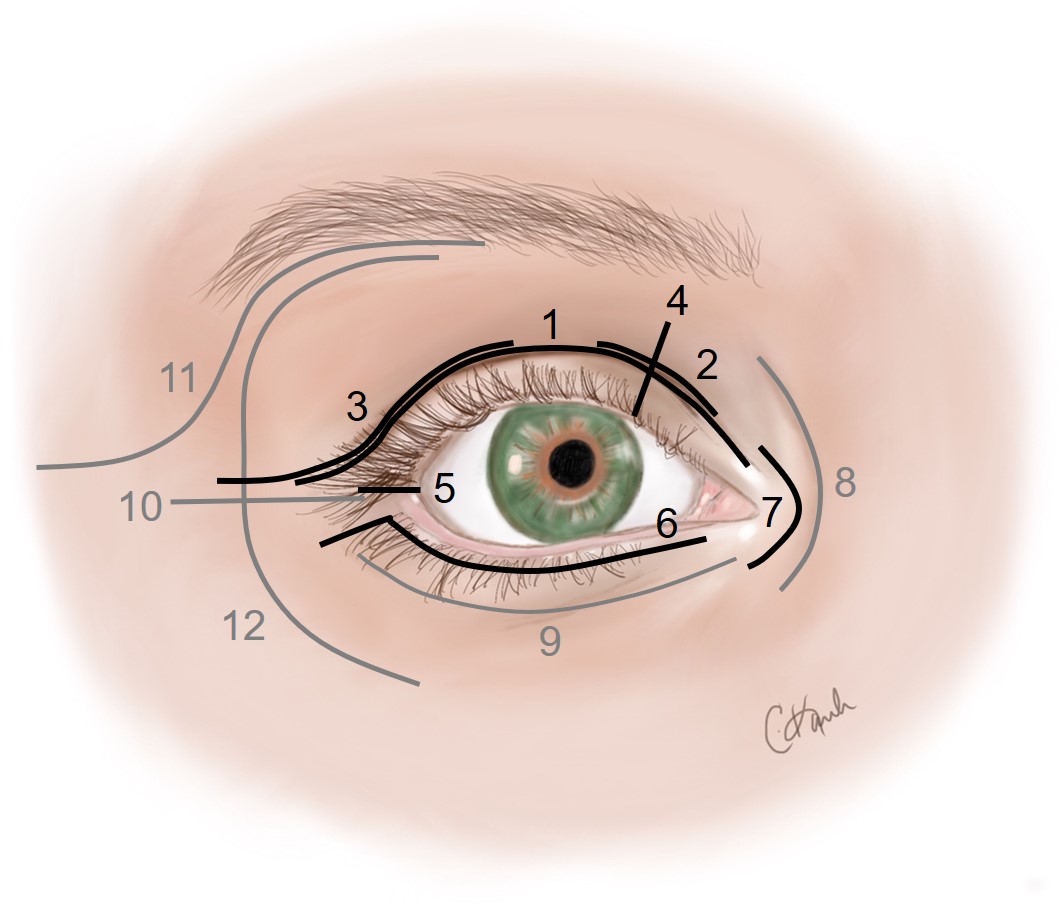
Figure 2. Illustration of various transcutaneous orbitotomy incisions, right eye. Incisions marked in gray are rarely utilized today. 1. Upper lid crease; 2. Medial lid crease; 3. Lateral lid crease; 4. Upper lid split; 5. Lateral canthotomy; 6. Subciliary; 7. Medial canthal tendon (Nunery); 8. Lynch; 9. Infratarsal; 10. Modified Berke; 11. Stallard-Wright; 12. Krönlein.
Transantral approach
- Access to orbital floor/medial wall for decompression (Walsh, Laryngoscope 1957)
- Technique:
- Upper gingivobuccal sulcus incision created
- Anterior wall of maxillary sinus removed
- Orbital floor/medial wall removed from below
- Maxillary antrostomy performed
- Mucosa closed with absorbable sutures
- Largely abandoned in favor of other approaches
Transnasal endoscopic approaches
- Endoscopic orbital decompression (Kennedy, Arch Otolaryngol Head Neck Surg 1990)
- Ethmoidectomy with removal of medial orbital wall +/- floor for thyroid orbitopathy or other congestive disorders
- Technique:
- Nasal cavity prepared with cottonoids soaked in oxymetolazone or epinephrine solution
- Medial turbinate medialized or removed as needed
- Complete ethmoidectomy performed
- Lamina papyracea removed
- Inferomedial strut and medial floor can also be removed
- Periorbita opened if desired
- Wide maxillary antrostomy performed
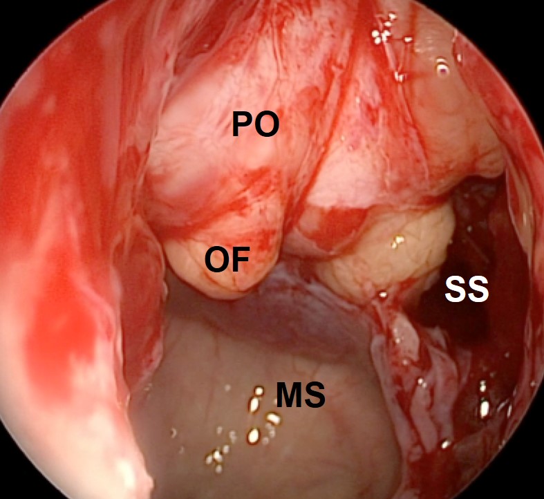
Figure 3. Intraoperative photograph of right endoscopic orbital decompression. The lamina papyracea and inferomedial strut have been removed, and the periorbita (PO) has been opened to facilitate prolapse of the orbital fat (OF). MS – maxillary sinus, SS – sphenoid sinus. Courtesy of Alexander Farag, MD, FARS.
- Endonasal medial orbitotomy (Yao, Curr Opin Otolaryngol Head Neck Surg 2016)
- Access to medial orbit/orbital apex for high-risk lesions which cannot be safely addressed from an anterior approach
- Technique:
- Ethmoidectomy performed as with decompression
- Limited removal of lamina papyracea, as much as is required to access lesion
- Periorbital opened sharply, blunt dissection carried into orbit to address lesion
- Bimanual approach can be facilitated by posterior septectomy and placement of second instrument through contralateral nares (Healy, OPRS 2014)
- If needed, medial wall can be reconstructed with nasal mucosal flap or alloplastic implant
- Optic canal decompression
- Removal of the medial wall of the optic canal for:
- Vision-threatening tumors of the sphenoid (Berhouma, Neurosurg Focus 2014)
- Traumatic optic neuropathy (benefit unproven) (Levin, Ophthalmology 1999)
- Technique:
- Endonasal ethmoidectomy performed
- Anterior wall of sphenoid sinus removed
- Medial wall of posterior orbit drilled to expose periorbita back to optic canal
- Bone over optic canal gently thinned to eggshell thickness with drill or ultrasonic aspirator
- Bone carefully elevated from nerve
- Removal of the medial wall of the optic canal for:
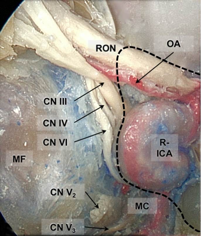
Figure 4.Endoscopic view of cadaver dissection of right orbital apex and sphenoid sinus. Extensive bone removal has been performed, including the sphenoid body, medial wall of the lesser sphenoid wing, ethmoid sinus and lamina papryacea, and optic strut. The sphenoid sinus is outlined by the dashed black line. RON – right optic nerve; OA – ophthalmic artery; R-ICA – right internal carotid artery (intracavernous portion); MC – Meckel’s cave; CN III/IV/V2/V3/VI – cranial nerves; MF – middle fossa. Courtesy of Alexandre Bossi Todeschini, MD.
Transorbital Neuroendoscopic Surgery
- Minimally invasive approach to anterior and middle cranial fossa (Moe, Neurosurgery, 2010)
- Access to
- Anterior cranial fossa
- Middle cranial fossa
- Cavernous sinus
- Meckel’s cave
- Set Up
- Brain lab navigation
- Four Handed Technique:
- Surgeon two hands available for instruments
- Assistant: holds the endoscope and orbital retractor
- Preferred technique to avoid prolonged compression on the globe or optic nerve
- Two Handed Technique
- Self-retaining retractor and scope holder
- Surgeon has two hands available for instruments
- Technique
- Lid crease incision lateral orbitotomy to allow exposure of the superior and inferior orbital fissure. A bone window may or may not be needed.
- To get to the anterior cranial fossa, the roof is drilled and the dura is opened
- To get to the middle cranial fossa, the deep portion of the greater wing of the sphenoid is drilled until the overlying dura is seen. A duroplasty is then performed.
- To see the cavernous sinus and Meckel’s cave, the meningoorbital band (most superficial dura band that tethers the front-temporal dura to the periorbita) must be cut
Transcranial approaches
- Minimally invasive subfrontal craniotomy (Reisch, Operative Neurosurgery 2005)
- Access to orbital roof for:
- Orbital roof fracture repair
- Orbital roof decompression
- Removal of lesions involving the anterior cranial fossa and superior orbit (e.g., meningioma, meningoencephalocele)
- Technique:
- Horizontal incision created through eyebrow
- Upper lid crease incision can also be used (Owusu Boahene, Skull Base 2010)
- Subperiosteal dissection performed over ipsilateral forehead, temporalis dissected
- Burr holes drilled, frontal craniotomy flap created (avoid frontal sinus)
- Frontal lobe dura dissected from floor of anterior cranial fossa
- Orbital roof removed as needed
- Bone flap/scalp flap replaced at case conclusion
- Access to orbital roof for:
- Frontotemporal craniotomy (Ringel, Neurosurgery 2007)
- Access to superior/lateral orbit for:
- Lesions of the sphenoid wing (e.g., meningioma)
- Large or high-risk lesions of the superior/lateral orbit
- Technique:
- Curvilinear scalp incision behind hairline from ear to contralateral forehead
- Temporalis muscle dissected from temporalis fossa
- Burr holes drilled, frontotemporal bone flap created, sparing orbital rim and zygomatic arch
- Dura over frontal and temporal lobes dissected from orbital roof and greater sphenoid wing
- Orbital roof and lateral wall removed with drill
- Following orbital surgery, orbital walls reconstructed as necessary
- Bone flap/scalp flap replaced at case conclusion
- Access to superior/lateral orbit for:
- Orbitozygomatic craniotomy (van Furth, Neurosurgery 2006)
- Access to superior/lateral orbit requiring greater exposure than that provided by frontotemporal approach
- Technique:
- Frontotemporal craniotomy performed as described above (without removing orbital roof/lateral wall)
- Subperiosteal dissection performed over superolateral orbital rim and zygomatic arch
- Osteotomies created through:
- Malar eminence
- Superolateral orbital rim
- Zygomatic arch
- Greater sphenoid wing (posterior lateral orbital wall)
- Superolateral orbit/zygoma removed en bloc
- Following orbital surgery, rim/zygoma replaced, orbital walls reconstructed as needed
- Bone flap/scalp flap replaced at case conclusion
- Transbasal (bilateral subfrontal) craniotomy (Feiz-Erfan, Skull Base 2008)
- Access to superior/medial/lateral orbit and sinonasal cavity for large or high-risk lesions of the orbit
- Technique:
- Bicoronal scalp flap created/dissected
- Temporalis muscle dissected bilaterally
- Bifrontal craniotomy performed
- Frontal sinus cranialized
- Supraorbital nerve released from notch/foramen
- Frontal bar removed
- Orbital roof(s) removed
- Following orbital surgery, orbital walls reconstructed as needed
- Bone flap/scalp flap replaced at case conclusion
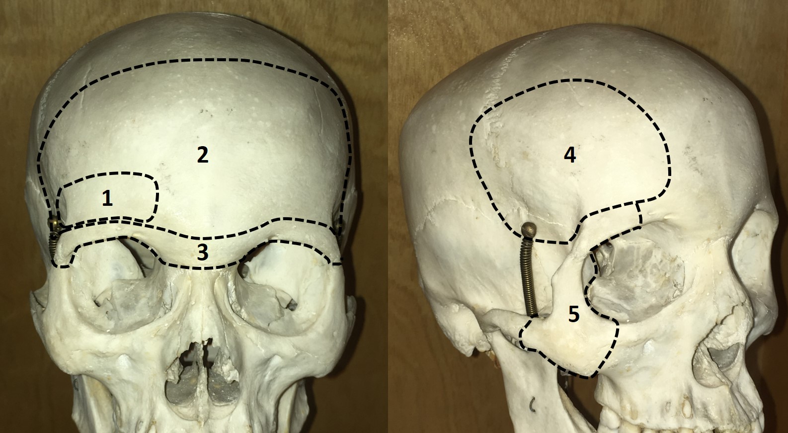
Figure 5.Craniotomy bone flaps used for transcranial orbitotomy. 1. Minimally invasive subfrontal; 2. Transbasal (bilateral subfrontal); 3. Frontal bar (often removed as part of transbasal approach); 4. Frontotemporal; 5. Orbitozygomatic (additional removal of orbital rim/zygoma in conjunction with frontotemporal flap.)
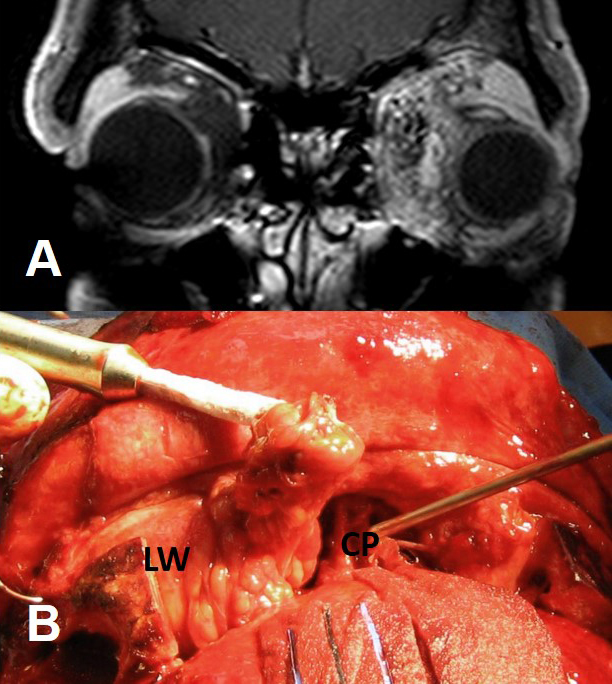
Figure 6. A, T1 fat-suppressed contrast-enhanced coronal MRI of 36 year old man with large left orbital arteriovenous malformation. B, Intraoperative photograph (superior surgeon’s view) showing bilateral subfrontal (transbasal) craniotomy approach to the left orbit. The frontal bar and orbital roof have been removed to expose the superior orbit. Cottonoids are covering the frontal dura and the tumor is being retracted with a cryoprobe. LW – lateral wall of the orbit, CP – cribriform plate.
Considerations in Pediatric Orbital Surgery
- Pediatric Orbital Anatomy (Tran, J Neurol Surg Skull Base, 2021)
- Neurocranial growth is continuous, with the greatest expansion by 2 years of age.
- At age 10, 95% of neurocranial growth has been achieved.
- Facial dimensions are 40% of the adult size by 3 months of age, and 80% by 5 years of age.
- Orbit reaches 75% of the adult depth by 6 years of age and 90% by 11 years of age.
- Sinus Development
- At birth maxillary sinus is underdeveloped and expands in biphasic growth at 3 and 7-18 years of age.
- The ethmoid and sphenoid sinuses grow in size between age 3 and 7.
- The paranasal sinuses develop to the front al sinus by 6 years of age.
- Pneumatization of the frontal sinus at the level of the orbital roof starts at 7 years of age and is complete by 15 years of age.
- Pneumatization of the sphenoid sinus is complete by 12 years of age.
- The same approaches of orbital surgery in an adult apply to pediatric population.
Patient management
- Postoperative instructions
- Activity restrictions
- Ice packs
- Elevate head of bed
- Avoid nose-blowing if sinuses entered
- Monitor for signs of bleeding or infection
- Postoperative expectations (swelling, bruising, diplopia, etc)
- Medications prescribed
- Topical antibiotics
- Pain medications
- Steroids if indicated
- Other management considerations
- Examine in recovery room to rule out catastrophic hemorrhage or visual dysfunction
- Manage in conjunction with ENT or neurosurgery in cases of endonasal or transcranial surgery
Treatment-related complications, prevention, and management
- Visual loss
- Incidence of severe vision loss 0.84% (Jacobs, Ophthalmology 2018)
- Highest with transcranial orbital roof surgery (18.2%), optic canal decompression (15.6%), and multiple facial fractures (6.5%)
- Etiology
- Orbital compartment syndrome
- Direct optic nerve/globe injury
- Damage to vascular supply to globe or optic nerve
- Posterior ischemic optic neuropathy (Rose, Orbit 2007)
- Prevention
- Intimate knowledge of orbital anatomy
- Meticulous surgical technique, avoiding excessive pressure on globe and tension on optic nerve
- Intraoperative pupil monitoring
- Avoidance of intraoperative hypotension
- Avoidance of epinephrine-containing solutions in orbit
- Management directed towards underlying etiology
- Orbital hemorrhage
- Signs/symptoms: severe pain, reduced vision, proptosis, ophthalmoplegia, hemorrhagic chemosis, increased intraocular pressure, relative afferent pupillary defect, funduscopic evidence of retinal vascular occlusion
- Prevention
- Preoperative cessation of anticoagulants
- Meticulous intraoperative hemostasis
- Management
- Immediate re-opening of wound with evacuation of hematoma
- Lateral canthotomy/cantholysis
- High dose corticosteroid therapy
- If no visual loss and pain is not severe, hemorrhage can be observed
- Diplopia
- Etiology
- Orbital edema/hemorrhage
- Damage to EOM
- Damage to nerve supply to EOM
- EOM entrapment or impingement by incompletely reduced fracture or malpositioned implant
- Herniation of orbital contents
- Postoperative scarring
- Prevention
- Knowledge of anatomy and proper surgical technique
- Management
- Treatment of underlying etiology
- Prisms
- Ocular occlusion
- Strabismus consultation with possible botulinum toxin therapy, strabismus surgery
- Globe malposition
- Enophthalmos, proptosis, hyper/hypoglobus, or medial/lateral displacement
- Etiology
- Incompletely reduced fracture
- Excessive decompression
- Improperly positioned implant
- Orbital fat atrophy (trauma, cautery, ischemia, radiation)
- Prevention
- Preoperative planning
- Proper implant sizing/shaping/positioning
- Meticulous surgical technique
- Management
- Treatment of underlying etiology
- Enophthalmos wedge
- Orbital volume augmentation (fat, fillers, etc)
- Eyelid malposition
- Ectropion, entropion, retraction
- Etiology
- Scarring of anterior lamella (ectropion) or posterior lamella (entropion) (Westfall, Ophthalmology 1991)
- Scarring or improper closure of septum (retraction)
- Prevention
- Proper incision placement
- Avoidance of excessive cautery to posterior/anterior lamella
- Judicious closure of incisions
- Management – treatment of underlying etiology
- Ptosis
- Etiology
- Damage to levator
- Damage to superior division of cranial nerve III
- Damage to Muller muscle/sympathetic nerve supply
- Mechanical displacement of levator (e.g., after orbital roof removal)
- Management
- Directed towards underlying etiology
- Typically observed for 6 months prior to surgical repair
- Neurotrophic keratopathy
- Nasociliary nerve damage
- Pupillary abnormalities
- Efferent pupillary defect due to ciliary ganglion trauma
- Loss of accommodation
- Dry eye
- Damage to lacrimal gland/ductules or lacrimal nerve
- Facial sensory loss
- Damage to branches of V1 or V2
- Cerebrospinal fluid leak
- Damage to dura beneath greater sphenoid wing, frontal bone, or cribriform plate
- 0.6-16% with exenteration, 1-3% with orbital decompression (Limawararut, Surv Ophthalmol 2008)
- Infection
Historical perspective
- Before the mid-20th century, most orbital surgery was performed by neurosurgeons or otolaryngologists (Alper, Doc Ophthal 1995)
- Krönlein (Swiss surgeon): lateral orbitotomy for removal of an orbital dermoid, 1888
- Dollinger (Hungarian surgeon): adopted Krönlein’s technique for lateral orbital decompression, 1911
- Lynch (New Orleans otolaryngologist): frontoethmoidal incision for frontal sinus fractures, 1921
- Hirsch (Viennese otolaryngologist): orbital floor decompression, 1929
- Naffziger (San Francisco neurosurgeon): orbital roof decompression, 1931
- Stallard (English ophthalmologist): modified Krönlein (Stallard-Wright) incision, 1946
- Walsh & Ogura (St. Louis otolaryngologists): transantral decompression, 1957
- Kennedy (Baltimore otolaryngologist): endoscopic orbital decompression, 1990
- In 1984, Maroon & Kennerdell reported using three different approaches to removing over 300 orbital tumors:
- Frontotemporal craniotomy
- Lateral orbitotomy (modified Berke incision)
- Medial peritomy with medial rectus disinsertion
- Today, oculoplastic surgeons perform the majority of orbitotomies through one of three approaches: (Reich, OPRS 2016)
- Transconjunctival inferior orbitotomy: McCord, 1979
- Upper lid crease incision: Harris, 1999
- Transcaruncular approach: Shorr, OPRS 2000
References and additional resources
- Alper MG. Pioneers in the history of orbital decompression for Graves’ ophthalmopathy. Doc Ophthal 1995;89:163-71.
- Berhouma M, Jacquession T, Abouaf L, et al. Endoscopic endonasal optic nerve and orbital apex decompression for nontraumatic optic neuropathy: surgical nuances and review of the literature. Neurosurg Focus 2014;37:E19.
- Bernardini FP, Nerad J, Fay A, et al. The revised direct transconjunctival approach to the orbital floor. Ophthal Plas Reconstr Surg 2017;33:93-100.
- Chabot JD, Gardner PA, Stefko ST, et al. Lateral orbitotomy approach for lesions of the middle fossa: a retrospective review of thirteen patients. Neurosurgery 2017;80:309-22.
- Esclamado RM, Cummings CW. Z-plasty modification of the Lynch incision. Laryngoscope 1989;99:986-7.
- Feiz-Erfan I, Spetzler RF, Horn EM, Porter RW, Beals SP, Lettieri SC, Joganic EF, DeMonte F. Proposed classification for the transbasal approach and its modifications. Skull Base 2008 Jan;18(1):29.
- Galbraith JEK, Sullivan JH. Decompression of the perioptic meninges for relief of papilledema. Am J Ophthalmol 1973;76:6887-92.
- Goldberg RA. Is there a “lost” rectus muscle in strabismus surgery? Am J Ophthalmol 2001;132:101-3.
- Hamed-Azzam S, Verity DH, Rose GE. Lateral canthotomy orbitotomy: a rapid approach to the orbit. Eye 2017;doi:10.1038/eye.2017.173.
- Harris GJ, Logani SC. Eyelid crease incision for lateral orbitotomy. Ophthal Plas Reconstr Surg 1999;15:9-18.
- Healy DY, Lee NG, Freitag SK, Bleier BS. Endoscopic bimanual approach to an intraconal cavernous hemangioma of the orbital apex with vascularized flap reconstruction. Ophthal Plas Reconstr Surg 2014;30:e104-6.
- Hill RH, Shiels WE, Foster JA, et al. Percutaneous drainage and ablation and first line therapy for macrocystic and microcystic orbital lymphatic malformations. Ophthal Plast Reconst Surg 2012;28:119-25.
- Jacobs SM, McInnis CP, Kapeles M, Chang SH. Incidence, risk factors, and management of blindness after orbital surgery. Ophthalmology 2018;125:1100-8.
- Kennedy DW, Goodstein ML, Miller NR, Zinreich SJ. Endoscopic transnasal orbital decompression. Arch Otolaryngol Head Neck Surg 1990;116:275-82.
- Kersten RC, Kulwin DR. Vertical lid split orbitotomy revisited. Ophthal Plas Reconstr Surg 1999;15:425-8.
- Krönlein RU. Zur pathologie und operative behandlung der dermoidcysten der orbita. Bruns Beitr Klin Chir 1888;4:149-63.
- Leone CR, Wissinger JP. Surgical approaches to diseases of the orbital apex. Ophthalmology 1988;95:391-7.
- Levin LA, Beck RW, Joseph MP, et al. The treatment of traumatic optic neuropathy: the International Optic Nerve Trauma Study. Ophthalmology 1999;106:1268-77.
- Limawararut V, Valenzuela AA, Sullivan, TJ, et al. Cerebrospinal fluid leaks in orbital and lacrimal surgery. Surv Ophthalmol 2008;53:274-84.
- Lynch RC. The technique of a radical frontal sinus operation which has given me the best results. Laryngoscope 1921;31:1-5.
- Maroon JC, Kennerdell JS. Surgical approaches to the orbit: indications and techniques. J Neurosurg 1984;60:1226-35.
- McCord CD, Moses JL. Exposure of the inferior orbit with fornix incision and lateral canthotomy. Ophthalmic Surg 1979;10:53-63.
- McKinney KA, Snyderman CH, Carrau RL, et al. Seeing the light: endoscopic endonasal intraconal orbital tumor surgery. Otolaryngol Head Neck Surg 2010;143:699-701.
- Miller NR, Monsein LH, Debrun GM, et al. Treatment of carotid-cavernous sinus fistulas using a superior ophthalmic vein approach. J Neurosurg 1995;83:838-42.
- Moe KS, Bergeron CM, Ellenbogen RG. Transorbital neuroendoscopic surgery. Neurosurgery. 2010 Sep;67(3 Suppl Operative):ons16-28.
- Naffziger HC. Progressive exophthalmos following thyroidectomy: its pathology and treatment. Ann Surg 1931;94:582-6.
- Owusu Boahene KD, Lim M, Chu E, Quinones-Hinojosa A. Transpalpebral orbitofrontal craniotomy: a minimally invasive approach to anterior cranial vault lesions. Skull Base 2010;20:237-44.
- Pelton RW, Patel BCK. Superomedial lid crease approach to the medial intraconal space: a new technique for access to the optic nerve and central space. Ophthal Plas Reconstr Surg 2001;17:241-53.
- Pelton RW. The anterior eyelid crease approach to the orbit. Curr Opin Ophthalmol 2009;20:401-5.
- Reich SS, Null RC, Timoney PJ, Sokol JA. Trends in orbital decompression techniques of surveyed American Society of Ophthalmic Plastic and Reconstructive Surgery members. Ophthal Plast Reconstr Surg 2016;32:434-7.
- Reisch R, Perneczky A. Ten-year experience with the supraorbital subfrontal approach through an eyebrow skin incision. Operative Neurosurgery 2005 Oct 1;57(suppl_4):ONS-242.
- Ringel F, Cedzich C, Schramm J. Microsurgical technique and results of a series of 63 spheno-orbital meningiomas. Neurosurgery 2007;60:214-21.
- Rose GE. The “devil’s touch”; visual loss and orbital surgery. A synopsis of the Mustardé Lecture, 2006. Orbit 2007;26:147-58.
- Rosen N, Priel A, Simon GJ, Rosner M. Cryo-assisted anterior approach for surgery of retroocular tumors avoids the need for lateral or transcranial orbitotomy in most cases. Acta Ophthalmol 2010;88:675-80.
- Shorr N, Baylis HI, Goldberg RA, Perry JD. Transcaruncular approach to the medial orbit and orbital apex. Ophthalmology 2000;107:1459-63.
- Stallard HB. Lateral orbitotomy (Krönlein’s operation). Br J Ophthalmol 1946;30:250.
- Timoney PJ, Sokol JA, Hauck MJ, Lee HB, Nunery WR. Transcutaneous medial canthal tendon incision to the medial orbit. Ophthal Plas Reconstr Surg 2012;28:140-4.
- Tran AQ, Kazim M. Orbital Surgical Guidelines: Pediatric Considerations. J Neurol Surg B Skull Base. 2021 Feb;82(1):142-148.
- van Furth WR, Agur AM, Woolridge N, Cusimano MD. The orbitozygomatic approach. Neurosurgery. 2006;58(1 Suppl):ONS103-107; discussion ONS103-107.
- Walsh TE, Ogura JH. Transantral orbital decompression for malignant exophthalmos. Laryngoscope 1957;67:544-68.
- Westfall CT, Shore JW, Nunery WR, et al. Operative complications of the transconjunctival inferior fornix approach. Ophthalmology 1991;98:1525-8.
- Yao WC, Bleier BS. Endoscopic management of orbital tumors. Curr Opin Otolaryngol Head Neck Surg. 2016;24:57-62.
