Upper Eyelid Blepharoplasty: Cosmetic and Functional Surgery
Updated January 2025
Peter J. Sneed, MD and Robert G. Fante, MD, FACS
Upper blepharoplasty is among the most common surgeries performed for the eyelids, and although it is fundamentally simple, there are many nuances and alternatives to optimize patient outcomes. Most patients will experience the benefit of both improved peripheral vision and improved appearance. This article reviews primary and revision surgery, and includes variations for phenotypic maintainance or alteration of East Asian eyelids.
Goals, indications, contraindications
Goals
- Restoration of normal function and appearance of the upper eyelids.
- Repair changes occur secondary to aging, hereditary features, inflammation, growth of abnormal tissue, or trauma.
- Improve visual function related to obstruction of the visual axis and/or obstruction to peripheral vision.
- Create or augment an upper eyelid crease.
- Improve preexisting asymmetry of crease position (or position of scar from prior surgery).
- Create or augment space for upper eyelid make-up.
- Improve appearance that can make patient feel more youthful.
- Help patient cope with difficult adjustments to change in appearance that may lead to anger, stress, anxiety, and depression.
- Avoid unrealistic expectations about change in appearance may limit patient acceptance of surgical result.
- Avoid unrealistic expectations that may also extend to anticipated improvement in quality of life.
Indications
- When excess upper eyelid skin obstructs vision, it affects daily activities.
- Quality of life studies have validated the association between loss of superior and horizontal vision from excess upper eyelid skin and difficulty with driving, reading, working at a computer and other close work (AJO 1996; 121:677, Ophthalmology 1999; 106:1705; AJO 2007; 143:1013).
- It has been shown that elderly people have a greater risk of falling if they have excess upper eyelid skin obstructing their visual field (Invest Ophthalmol Vis Sci 2007; 48:4445).
- Patients often complain of headache and brow ache from overworked frontalis muscles, employed to lift excess skin up from the eyelid margins.
- Skin lying on the eyelashes — produces discomfort independent of obstructed visual axis.
- Dermatitis — chronic dermatitis caused by redundant skin is an indication for surgery.
- Patients undergo upper blepharoplasty for purely aesthetic reasons, including multiple creases, skin crepiness, heaviness of eyelid folds, eyelid crease asymmetries, eyelash ptosis, epicanthal fold reduction, scar camouflage, xanthelasma removal, gender alteration, and more.
Contraindications
All relative:
- Dry eyes
- Compromised blink
- Cicatricial or paralytic lagophthalmos
- Unrealistic expectations or Body Dysmorphic Disorder
- Anticoagulants may increase the risk of post-operative bleeding.
- Aspirin products – Ecotrin®, Fiorinal®, Percodan®
- Non-steroidal anti-inflammatory drugs – ibuprofen, naproxen, piroxicam
- Nutritional supplements – fish oil, vitamin E, gingko biloba, ginseng
- Warfarin, Apixaban (Eliquis®), Dabigatran (Pradaxa®), Clopidogrel (Plavix®), Acetylsalicylic acid/Dipyridamole (Aggrenox®)
- Cold urticaria or history of hives, anaphylaxis, or swelling after contact with cold objects may cause increased swelling post operatively
Preprocedure evaluation
Patient history
- Explain and document how daily visual function is affected.
- Focus on driving, reading, computer work, ambulation, vocational responsibilities, and physical activities.
- Loss of peripheral vision
- A questionnaire for the patient to complete may be helpful for insurance or Medicare preauthorization (see Appendix A).
- Forehead aches from chronic lifting of eyebrows, and consequent exacerbation of visual obstruction at the end of the day or when tired.
- History of dry eyes and current treatment protocol
- Daytime and Nighttime artificial tears or ointments
- Treatment of Meibomian Gland Disease (MGD)
- Prior LASIK or other refractive surgery
- Cyclosporine A (Restasis®), Lifitigrast (Xiidra®), serum tears
- History of facial paralysis and/or infrequent blinking
- Previous ocular surgeries
- Past ocular history
- Aesthetic wishes
- Expectations of the surgical procedure
- Significant life changes or stresses.
Clinical examination
External examination:
- Special attention to quality, quantity, and symmetry of the eyelid skin.
- Unilateral or bilateral eyebrow ptosis and/or frontalis compensation (often unrecognized by the patient).
- Eyelid crease: Presence or absence, height and number
- For both functional and cosmetic East Asian patients, discussion of the patient’s desired eyelid configuration is important. Single or double eyelid configurations may be appropriate based on patient preferences.
- Upper and lower eyelid margin position.
- Upper eyelid excursion
- Lid closure and seventh nerve function.
- Presence or absence of Bell phenomenon.
- Presence and degree of severity of floppy eyelid syndrome
- Scars from trauma or previous surgery
- Note any resistance to passive lid movement
Slit Lamp Examination
- Tear film quality and blink mechanism. In patients with history or clinical signs of dry eye, consider basal tear secretion testing, or Schirmer II with anesthesia testing.
- Eyelid margin disease, including MGD, trichiasis, blepharitis
- Corneal and conjunctival examination and documentation of any disease
Preoperative assessment
- Measurement of margin reflex distance (MRD)
- Palpebral fissure distance in primary and downgaze (PF)
- Levator function (LF)
- Assess nasal fat pad and preaponeurotic fat pad protrusion.
- Assess presence and/or degree of lacrimal gland prolapse.
- Measure skin amount in millimeters between the lower border of the central brow and the eyelash margin.
- Visual field testing with eyebrows relaxed, patient looking straight ahead, and the eyelids in normal relaxed position
- Visual field is repeated with the eyelids taped up.
- Photographs of frontal plane and oblique view.
- Advantages:
- Flash photography documents the MRD and corneal light reflex as well any eyelid skin resting on the eyelashes.
- Document preoperative eyelid and facial abnormalities or asymmetries.
- Help explain to the patient unique facial features important for planned surgical procedure.
- Preoperative to postoperative comparisons illustrate surgical changes.
- Part of the medical record (medicolegal and insurance-related issues).
- Insurance plans may require additional photographs with the eyebrows and excess eyelid skin taped upward.
- If brow ptosis is present, frontal photographs often demonstrate involuntary frontalis muscle compensation.
- Advantages:
- For functional surgery depending on anticipated insurance or Medicare coverage (carrier specific).
- Visual field testing with eyebrows relaxed, patient looking straight ahead and the eyelids in normal relaxed position.
- Visual field testing repeated with the eyebrows and excess eyelid skin taped up.
Procedure alternatives
- Alternative treatments should be always be reviewed as part of the informed consent discussion with the patient.
- Blepharoptosis repair (if present)
- Forehead/Eyebrow Lift
- When significant eyebrow ptosis is present, reconstructive lift of the eyebrows may be appropriate concomitantly with upper blepharoplasty or as an alternative.
- For cosmetic patients, eyebrow lift and/or eyebrow fat pad volumization may also help achieve their aesthetic goals.
- Concomitantly with upper blepharoplasty or as a stand-alone procedure.
- Vrcek I, Chou E, Somogyi M, Shore JW. Volumization of the brow at the time of blepharoplasty: treating the eyebrow fat pad as an independent unit. Ophth Plast Reconstr Surg 2018;34(3):209-212.
- Trepsat F. Periorbital rejuvenation combining fat grafting and blepharoplasties. Aesth Plast Surg 2003;27:243-253.
- Controversy exists on whether blepharoplasty affects eyebrow position:
- No effect on brow position
- Dar SA, Rubinstein TJ, Perry JD. Eyebrow position following upper blepharoplasty. Orbit 2015;34(6):327-30.
- Nakra T, Modjtahedi S, Vrcek I, et al. Effect of Upper Eyelid Blepharoplasty on Eyelid and Brow Position. Orbit 2016;35(6):324-327.
- Brow position may be lower when frontalis recruitment is no longer necessary
- For everyone: Prado RB, Silva-Junior DE, Padovani CR, Schellini SA. Assessment of eyebrow position before and after upper eyelid blepharoplasty. Orbit 2012;31(4):222-6.
- For men only: Huijing MA, van der Palen J, van der Lei B. Effect of upper eyelid blepharoplasty on eyebrow position. J Plast Reconstr Aesthet Surg 2014;67(9):1242-7.
- For patients with co-existing blepharoptosis only:
- Lee JM, Lee TE, Lee H, et al. Change in brow position after upper blepharoplasty or levator advancement. J Craniofac Surg 2012;23(2):434-6.
- Rootman DB, Karlin J, Moore G. Goldberg R. Effect of ptosis surgery on brow position and the utility of preoperative phenylephrine testing. Ophth Plast Reconstr Surg 2016;32(3):195-8.
- No effect on brow position
- Relative merits and disadvantages of addressing concurrent blepharoptosis, eyebrow ptosis, eyelid retraction, and other sources of eyelid, eyebrow and orbital asymmetry should be discussed during surgical planning.
- Racial and ethnic facial characteristics including skin type and underlying facial bone structure may be included in discussing alternatives and surgical planning.
- Pre-operative preparation may include asking the patient to stop smoking, reduce alcohol intake, and optimize overall general health.
Surgical techniques
Skin marking is influenced by gender, race, and unique facial features of each patient.
- Gender
- In women, the brow and lid creases are higher and more arched, and the lid fold is less prominent.
- In men, the brow protrudes more anteriorly, and is generally flatter.
- Race
- Typically, the eyelid crease lies 4–12 mm above the lash line.
- In Caucasian and African-American women, the crease is usually 8-11 mm above the lid margin.
- In Caucasian and African-American men, the crease is usually 6-9 mm above the eyelid margin.
- The East Asian upper eyelid has more fullness, narrower palpebral fissures, more prominent medial epicanthal folds, and a lid crease closer to the eyelid margin.
- Lid crease in East Asians can be absent, nasally tapered, or flat but typically lies lower (4-7 mm above the lid margin or may be as low as 1 mm above the eyelashes when there appears to be no crease at all) and flatter than Caucasians.
- In Caucasians, the orbital septum attaches to the levator aponeurosis at or slightly above the superior tarsal border or over the anterior surface of the tarsus.
- In East Asians, the orbital septum fuses to the levator aponeurosis at variable distances below the superior tarsal border.
- Preaponeurotic fat pad protrusion and a thick subcutaneous fat layer prevent levator fibers from extending toward the skin near the superior tarsal border.
- The primary insertion of the levator aponeurosis into the orbicularis muscle and into the upper eyelid skin occurs closer to the eyelid margin in East Asians.
- The East Asian eyelid includes some pretarsal fat pad and may include more volume in the preaponeurotic fat pads.
- Typically, the eyelid crease lies 4–12 mm above the lash line.
- Unique facial features
- Patients may prefer to retain or change certain features such as relative hollowness or fullness of the upper eyelid sulcus. Removal or preservation of fat and muscle can help achieve these goals. Review of old or family photographs may be helpful in clarifying preferences and objectives.
- Skin Marking
- One approach to assuring that sufficient skin remains for complete closure of the eyelid is to leave at least 20mm of skin in the eyelid after surgery.
- A total of 20 mm of skin should remain when measured vertically between the lower margin of the central eyebrow and the margin of the central eyelashes.
- For example, if the lid crease is marked 8 mm above the lash margin, the upper edge of the incision should be 12 mm below the brow margin.
- An alternative approach is the “pinch method” where eyelid skin is grasped and gathered until the skin is tight and the lashes begin to evert.
- The lower lateral marking is extended to the orbital rim or end of the eyebrow and may course superiorly or follow existing creases to meet the upper mark.
- Extending the marking too far lateral may result in unwanted visible scarring.
- However with skin closure, this scar generally blends well with the normal smile lines in the lateral canthal area.
Variations for East Asians
- Trapezoidal upper blepharoplasty for double eyelid creation
- One method to create a stable double eyelid: a narrow ellipse of skin with a wider ellipse of orbicularis can be excised (a trapezoid in cross-section) to permit strong attachment to the levator aponeurosis. Fat is typically not removed. (See Figure 1)
- Jang JW. Double-eyelid surgery: incisional techniques. In: Jin HR ed. Aesthetic Plastic Surgery of the East Asian Face. New York, NY: Thieme; 2016:162–172.
- Chen WPD, ed. Asian Blepharoplasty and the Eyelid Crease, 3rd ed. Philadelphia, PA: Elsevier; 2016:169–181.
- Medial epicanthoplasty
- Several techniques depending on type of epicanthal fold including modified Uchida W-plasty, Parks Z-plasty, Root Z-plasty, and redraping epicanthoplasty.
- The epicanthal incisions should not connect to the blepharoplasty incision.
- Extremely delicate, tension-free closure is necessary to avoid scarring.
- Baek JS, Choi YJ, Jang JW. Medial epicanthoplasty: what works and what does not. Facial Plast Surg 2020;36:584-591.
Injection of local anesthetic
- Timing
- May be administered in the operating room or preoperative holding area.
- Early injection takes advantage of the time required to move, position, prep, and drape the patient, during which time the anesthetic will take effect.
- Dosing
- 1% or 2% lidocaine with 1:100,000–200,000 units of epinephrine is typically used, sometimes with the addition of hyaluronidase.
- Approximately 1–1.5 cc of anesthetic is injected through a 27- or 30-gauge needle in the plane between skin and orbicularis muscle across the entire eyelid.
- Great care is taken to point the needle away from the globe, to avoid inadvertent penetration with sudden patient movement.
- In lidocaine (amide-type) sensitive patients, procaine (ester-type) may be used.
- The addition of epinephrine to local anesthetic solutions prolongs the duration of action of the anesthetic agent and may reduce intraoperative bleeding.
Tissue excision
- Some surgeons prefer to place a corneal protector in each eye.
- This is particularly important if incisions are made with the CO2 laser.
- The incision, which is made along the previously marked lines, can be made with a 15 Bard Parker blade, an incisional CO2 laser, a diamond blade, or a needle-tipped Bovie or radiofrequency instrument. The collateral damage caused by laser or cutting cautery may induce incisional inflammation, especially in more sebaceous skin sometimes seen in East Asians; scalpel incision may be preferable.
- Depth of excision depends on the preoperative plan.
- Excess skin only is typically be removed.
- Orbicularis and/or fat may be removed in addition.
- Partial removal of orbicularis muscle over the lateral eyelid area with grafting of medial fat into the lateral sub-brow area has been proposed to restore youthful contours (Fezza J, OPRS 2012;28:446).
- The tissue to be excised is grasped with a forceps and meticulously dissected along the intended plane.
- Cautery is applied for hemostasis.
- Excess central preaponeurotic and/or nasal fat is removed.
- Care is taken not to remove excessive fat, particularly in the pupillary meridian where inadequate volume can lead to an A frame deformity.
- The whiter nasal fat is more commonly prolapsed and can be removed or transposed. If some nasal fat is to be removed, care is taken to cauterize or avoid medial palpebral vessels which course over the medial fat pad to prevent deep vascular trauma and subsequent retrobulbar hemorrhage. (Callahan MA. Prevention of blindness after blepharoplasty. Ophthalmology 1983;90(9):1047-51.)
- Gentle cautery applied to the orbital fat may contour and reposition the remaining fat posteriorly into the orbit, providing needed volume and fullness.
- Care is taken to avoid injury to the levator palpebrae superioris complex which lies just posterior to the preaponeurotic fat pad.
Wound closure
- In addition to primary closure of the skin, attention may focus on creation of symmetric and well-positioned eyelid creases.
- Lid crease fixation is not always necessary, especially for men.
- When needed, lid crease fixation method depends on surgeon’s preferences and experience.
- Interrupted suture placement can incorporate superficial fibers of levator aponeurosis just above the superior edge of the tarsal plate. The tarsus itself should not be used for lid crease fixation since it will cause an abnormal crease that persists with eyelid closure.
- Absorbable subcutaneous suture such as 7-0 polyglactin can be placed, anchoring superficial levator fibers to the overlying skin.
- Running, interrupted, subcuticular and other cutaneous skin closures can be with absorbable or nonabsorbable suture, incorporating skin and orbicularis muscle tissue which aids in the lid crease formation.
- As an alternative to suture closure, some surgeons prefer octyl-2-cyanoacrylate for blepharoplasty wound closure.
- Temporary sutures may approximate the skin before application of the glue.
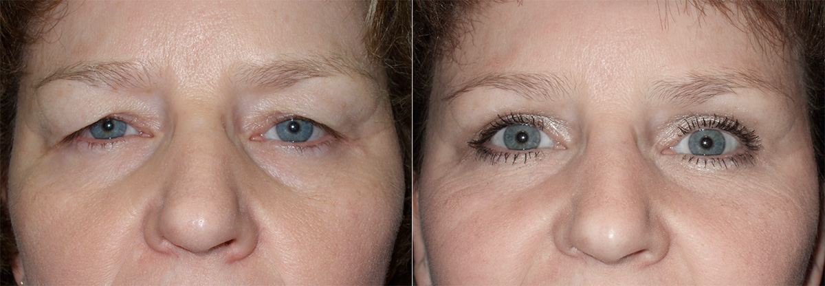
Figure 1 – before and after upper trapezoidal blepharoplasty to create a double-eyelid in an East Asian (courtesy of Robert Fante, MD).

Figure 2 – before and after upper blepharoplasty in a Caucasian using lid crease fixation (courtesy of Robert Fante, MD).


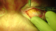
Video 1. Laser can be used to expose the superficial fibers of the levator for incorporation into the skin closure to augment the eyelid crease.

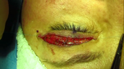
Video 2. Subcuticular closure.
Wound care
- Antibiotic ointment may be placed over incision.
- Moistened gauze may be placed over the closed eyelids.
- Many surgeons apply a cold compress while the patient is in the recovery area.
- Before discharge, wounds are checked for bleeding and dehiscence.
Ongoing controversies
As noted above in Alternatives to the Procedure, the effect of blepharoplasty on eyebrow position is controversial.
Patient management: treatment and follow-up
Postoperative instructions
- Patients may usually resume most normal activities within 24-48 hours after surgery, although many surgeons recommend limiting physical exertion for 7-14 days.
- Discomfort and edema are expected after surgery and are usually adequately managed with acetaminophen.
- Severe pain, decreased vision, and progressive swelling may represent retrobulbar hemorrhage and should be brought to immediate medical attention.
- Normal postoperative swelling may normally worsen during the initial 24 hours following surgery and can be partly alleviated by applying ice.
- Degree of swelling is related to surgical factors such as ecchymosis, cauterization, tissue manipulation, and patient response to surgery.
- Dissection in the lateral canthal area may result in altered lymphatic drainage.
- Head elevation and limiting activity may reduce edema.
- Progressive postoperative periorbital inflammation may indicate infection, allergy to topical medication and rarely primary acquired cold urticaria (PACU).
- Patients who experience severe itching, erythema, and progressive conjunctival injection should be advised to discontinue topical ointment due to possible allergy.
- Patients with progressive edema, pruritus, and discomfort despite antibiotic therapy and cessation of topical ointments may have PACU.
- A cold stimulation test may confirm the diagnosis of PACU.
- Patient education and cold avoidance are the primary means of treatment.
- Nonsedating antihistamines may help control cold-induced symptoms.
- An allergist should guide the workup and management of this condition.
- Patients with previously established PACU can still undergo surgery if appropriate safety precautions are followed.
Medications prescribed
- Antibiotic or steroid/antibiotic ointment may be applied twice a day to sutures and into the eyes at night.
- Artificial tears may also be recommended.
Other management considerations
- Bruising and swelling typically lasts 10–14 days after surgery, but final results are best appreciated at ~6 weeks. Final scarring can take up to a year.
- Frequency of cold compresses is decreased as the effectiveness of this therapy lessens.
- Contact lens wear may be resumed at approximately 1 week postop, but patients should insert and remove contact lenses by manipulating the lower eyelid in order to prevent wound dehiscence. This is especially relevant at the vulnerable lateral canthal area.
- Homeopathic treatments such as Bromelain and Arnica montana are sometimes suggested to minimize post-operative bruising and swelling. However, Level 1 scientific evidence has failed to find a beneficial effect from these treatments.
- Kotlus BS, et al. Evaluation of homeopathic Arnica montana for ecchymosis after upper blepharoplasty: a placebo-controlled, randomized, double-blind study. Ophth Plast Reconstr Surg 2010;26(6):395-7.
- Van Exsel DCE, et al. Arnica ointment 10% does not improve upper blepharoplasty outcome: a randomized, placebo-controlled trial. Plast Reconstr Surg 2016;138(1):66-73.
- Nonabsorbable sutures are removed 7-14 days after surgery.
- Absorbable sutures vary in rate of absorption and degree of inflammation – often they are removed as well.
- Postoperative eyelid numbness involving the upper eyelid skin and eyelashes is an expected outcome after upper blepharoplasty and typically resolves over 2 to 4 months.
- Sensory nerve fibers from the supraorbital, supratrochlear, and lacrimal nerves travel in the preorbicularis plane, suborbicularis fascial plane, and within the orbicularis. These distal branches of the ophthalmic division of the trigeminal nerve are transected during supratarsal eyelid crease incision for blepharoplasty and ptosis repair.
- Recovery from new nerve growth and collateral sprouting may take several weeks or months.
Preventing and managing treatment complications
Patient dissatisfaction
- Unfortunately, even with careful patient selection and surgical planning, and an uneventful perioperative period, some patients may be dissatisfied with their results.
- Establishing a good patient-surgeon bond preoperatively is essential to managing any real or perceived surgical complication that may occur.
- Patients with unrealistic expectations may perceive an operative complication after uncomplicated surgery.
Wound dehiscence
- Wound dehiscence may be due to inadvertent trauma, poor wound healing, excessive tension, early suture removal, infection, hematoma, and seroma formation.
- Patients may inadvertently rub their eyes in the hours after surgery when their lids are numb or while sleeping.
- Often this occurs laterally where there is increased vertical tension.
- Interrupted sutures are used to reapproximate the wound edges. Wound edges are freshened, and granulation tissue removed.
- Smaller areas of dehiscence typically self-resolve seamlessly, though final results may only be known several weeks later. Closer follow-up is recommended when allowing for secondary intention healing, and patients should be reassured that later touch-up can be performed.
Exacerbation of dry eye syndrome
- Careful preop evaluation and perioperative artificial tears, ointments, punctal plugs can help prevent worsening of dry eyes.
Scar hypertrophy
- Hypertrophic scarring is uncommon in the thin eyelid skin. Keloids at the upper eyelids have never been reported in the medical literature.
- Treatment includes vitamin E cream, massage, and topical or injected corticosteroids.
Suture granuloma
- More frequent with absorbable sutures and may be removed or treated with steroid injections.
Inclusion cyst
- Sequestered epithelial remnants along the suture line.
- Usually atrophy in 2 to 3 months.
- May be managed by rupturing the cyst and marsupialization with an 18-gauge needle.
Overcorrection and lagophthalmos
- May occur if too much skin is removed.
- Usually preventable with the 20-mm rule described above.
- Tension in the levator complex and orbital septum may also result in eyelid retraction. Care should be taken during suture placement that the orbital septum is not included in the skin closure.
- Excessive skin removal may require free full-thickness skin grafting.
- Lagophthalmos due to internal scarring requires surgical exploration and scar lysis.
- Occasionally spacer grafts are required to completely correct the lid retraction.
Undercorrection
- After at least 3-6 months healing, persistent excess skin and/or fat-related fullness can be simply treated with a revision upper blepharoplasty.
Excessive orbital fat removal
- May result in a hollowed appearance.
- May require micro fat grafting, dermis-fat graft, or filler injection to correct the upper eyelid volume deficiency.
Asymmetry
- May be related to surgery or preoperative asymmetry of the face, lid, or brow.
- Lid crease asymmetry is usually corrected by raising the lower eyelid crease.
- Lowering a high lid crease has a lower success rate.
- May be accomplished by securing posterior skin to the levator complex at the superior border of the tarsal plate.
- Lateral skin often takes longer to soften and smooth because it is thicker compared to eyelid skin.
Medial canthal webbing
- May occur when the incision extends too far medially.
- Prevent by planning an incision that extends to the medial commissure.
- May be corrected by Z-plasty, W-plasty, transposition flaps, or Y-V advancement procedures.
Ptosis
- May be due to inadvertent trauma to the levator complex, including postsurgical edema and dehiscence.
- May be due to unrecognized preoperative levator dehiscence.
Epithelial keratopathy
- May be related to lagophthalmos and dry eye.
- Usually corrected with lubrication regimen.
- May require corrective lid surgery to reduce palpebral aperture.
Epiphora
- May be related to corneal irritation and/or dryness.
- Dry eye symptoms may worsen if there is a decreased blink with orbicularis excision.
- Inadvertent injury to the lacrimal system should be avoided during upper blepharoplasty by limiting the incision medially.
Diplopia
- Rare
- May be from diffusion of local anesthetic affecting one or more extraocular muscles. Should resolve within 24 hours.
- Inadvertent trauma to an extraocular muscle with deep dissection in orbital fat.
- Cautery may affect nerve or muscle.
- The superior oblique muscle and trochlea are vulnerable to surgical trauma because of their anterior position in the orbit (Plast Reconstr Surg 2001;108:2137).
Globe injury
- May occur with CO2 laser, steel scalpel, radiofrequency needle, or local anesthetic injection
- When CO2 laser is used, protective corneal shields are imperative. Laser is always directed away from the globe when cutting.
Orbital hemorrhage and blindness
The most serious complication following upper blepharoplasty, and the most common cause for large indemnity awards for medical malpractice cases involving ophthalmologists and eyelid surgery (Fante RG et al. Medical professional liability claims: experience in oculofacial plastic surgery. Ophthalmology 2018;125(12):1996-1998).
- Rare, with an estimated incidence of 1:20,000 (Ophthal Surg 1990;21:85).
- Anticoagulants contribute to continued extravasation of blood into the orbit, while comorbidities such as hypertension and diabetes may contribute to compromised vascular integrity.
- Prolonged surgery and reoperation with scarred tissue contribute to swelling and ecchymosis.
- Acute orbital hemorrhage requires prompt intervention.
- Postoperative patches and bandages are removed in the recovery room to permit early detection of postoperative bleeding.
- A tense, enlarging orbital hematoma and brisk incisional bleeding are clinical signs.
- Proptosis, severe pain, decreased visual acuity, relative afferent pupillary defect, and elevated intraocular pressure confirm the diagnosis.
- Retrobulbar hemorrhage is a form of compartment syndrome, with pressure rising abruptly within the fixed walls of the orbit.
- Blood supply to critical structures including the optic nerve become compromised. Rapid treatment is critical.
- Treatment is focused partly on identifying the source of bleeding, but frequently, active bleeding subsides from tamponade within the closed orbital compartment.
- Rapid release of orbital pressure by opening the wound, releasing the lid with a lateral canthotomy with inferior and/or superior cantholysis, is most important.
- Prompt decompression of the orbit alone can restore vision.
- The wound may be left open or closed loosely.
- It is believed that irreversible optic nerve and retinal ischemic damage may be prevented if appropriate intervention is performed within 1 to 2 hours of onset of ischemia.
- If canthotomies have not restored vision, spreading bluntly posteriorly into the orbit along the lateral wall to access and release deep hematomas may be helpful.
- Systemic osmotic agents and corticosteroids may be given but do not take the place of prompt pressure release.
- IV acetazolamide (500 mg)
- IV mannitol 20% (1–2 g/kg over 30–60 minutes)
- IV methylprednisolone 100 mg
- Involvement of an internist or hospitalist is helpful in managing fluid shifts caused by these osmotic agents.
- Rarely, bony decompression, either at bedside through the inferomedial floor or in the operating room, can be considered.
- CT scan is important, but only after initial decompression treatment has been completed.
- Post-treatment admission to hospital is recommended, with close visual acuity monitoring, head elevation, ice water compresses, and intravenous steroids until 24 hours of stable vision have been noted.
- Topical and systemic antibiotics are given for the open wounds.
- Wounds may be repaired electively in 1 to 2 weeks not closing well by secondary
Common treatment responses, follow-up strategies
- Upper blepharoplasty can yield significant functional and aesthetic benefits for patients.
- Identifying patients with body dysmorphic syndrome, dysmorphophobia, or narcissistic behavior helps screen for those who may not be appropriate candidates.
- Even well-adjusted patients will perceive and focus on asymmetry caused by bruising and swelling or discomfort during the early postoperative period.
- Secondary revision surgery should remain an option during follow-up treatment and should be considered normal. Typically, this occurs weeks to months after the initial surgery.
History
- Historically the term blepharoplasty has had a broader context.
- Aulus Cornelius Celsus was a first-century Roman who described making an incision in the skin to relax the eyelids (Orbit 2012;31:162).
- In the tenth century, Middle Eastern surgeons described removal of excess eyelid skin to improve vision. It was then used by Karl Ferdinand von Graefe in 1818 when describing eyelid repair after removal of skin cancer (Plast Reconstr Surg 1971;47:246).
References and additional resources
- Adams J, Murray R. The general approach to the difficult patient. Emerg Med Clin North Am 1998; 16:689.
- Battu VK, Meyer DR, Wobig JL. Improvement in subjective visual function and quality of life outcome measures after blepharoptosis surgery. Am J Ophthalmol 1996;121:677.
- Baek JS, Choi YJ, Jang JW. Medial epicanthoplasty: what works and what does not. Facial Plast Surg 2020;36:584-591.
- Black EH, Gladstone GJ, Nesi FA. Eyelid sensation after supratarsal lid crease incision. Ophthal Plast Reconstr Surg 2002; 18:45.
- Brown MS, Siegel IM, Lisman RD. Prospective analysis of changes in corneal topography after upper eyelid surgery. Ophthal Plast Reconstr Surg 1999;15:378.
- Burnstine MA, Putterman AM. Upper blepharoplasty: a novel approach to improving progressive myopathic blepharoptosis. Ophthalmology 1999;106(11):2098-100.
- Burroughs JR, Patrinely JR, Nugent JS, et al: Soparkar CNS, Anderson RL, Pennington J H. Cold urticaria: an underrecognized cause of postsurgical periorbital swelling. Ophthal Plast Reconstr Surg. 2005; 21:327.
- Callahan MA. Prevention of blindness after blepharoplasty. Ophthalmology 1983;90(9):1047-51.
- Chen WPD, ed. Asian Blepharoplasty and the Eyelid Crease, 3rd ed. Philadelphia, PA: Elsevier; 2016:169–181.
- Dar SA, Rubinstein TJ, Perry JD. Eyebrow position following upper blepharoplasty. Orbit 2015;34(6):327-30.
- Dupuis C, Rees TD: Historical notes on blepharoplasty. Plast Reconstr Surg 1971; 47: 246.
- Fante RG, Bucsi R, Wynkoop K. Medical professional liability claims: experience in oculofacial plastic surgery. Ophthalmology 2018;125(12):1996-1998
- Federici TJ, Meyer DR, Lininger LL. Correlation of the vision-related functional impairment associated with blepharoptosis and the impact of blepharoptosis surgery. Ophthalmology 1999; 106:1705.
- Freeman EE, Muñoz B, Rubin G, West SK. Visual field loss increases the risk of falls in older adults: the Salisbury Eye Evaluation. Invest Ophthalmol Vis Sci 2007; 48:4445.
- Goldberg RA, Marmor MF, Shorr N, Christenbury JD. Blindness following blepharoplasty: two case reports, and a discussion of management. Ophthalmic Surg 1990; 21:85.
- Hass AN, Penne RB, Stefanyszyn MA, Flanagan JC. Incidence of postblepharoplasty orbital hemorrhage and associated visual loss. Ophthal Plast Reconstr Surg 2004; 20:426.
- Heinze JB, Hueston JT. Blindness after blepharoplasty: mechanism and early reversal. Plast Reconstr Surg 1978; 61:347.
- Huijing MA, van der Palen J, van der Lei B. Effect of upper eyelid blepharoplasty on eyebrow position. J Plast Reconstr Aesthet Surg 2014;67(9):1242-7.
- Jang JW. Double-eyelid surgery: incisional techniques. In: Jin HR ed. Aesthetic Plastic Surgery of the East Asian Face. New York, NY: Thieme; 2016:162–172.
- Jeong S, Lemke BN, Dortzbach RK, et al: The Asian upper eyelid: an anatomical study with comparison to the Caucasian eyelid. Arch Ophthalmol 1999; 117:907.
- Jordan DR, Mawn LA. Dysmorphophobia. Can J Ophthalmol 2003; 38:223.
- Kotlus BS, et al. Evaluation of homeopathic Arnica montana for ecchymosis after upper blepharoplasty: a placebo-controlled, randomized, double-blind study. Ophth Plast Reconstr Surg 2010;26(6):395-7.
- Lazzeri D, Agostini T, Figus M et al: The contribution of Aulus Cornelius Celsus (25 B.C.-50 A.D.) to eyelid surgery. Orbit 2012; 31:162.
- Lee CW, Sheffer AL. Primary acquired cold urticaria. Allergy Asthma Proc 2003; 24:9.
- Lee JM, Lee TE, Lee H, et al. Change in brow position after upper blepharoplasty or levator advancement. J Craniofac Surg 2012;23(2):434-6.
- Lelli GJ, Lisman RD: Blepharoplasty complications. Plast Reconstr Surg 2010; 125:1017.
- Lewis CM, Lavell S, Simpson MF. Patient selection and patient satisfaction. Clin Plast Surg 1983; 10:321.
- Mackley CL. Body dysmorphic disorder. Dermatol Surg 2005; 31:553.
- McCullough ME, Emmons RA, Kilpatrick SD, Mooney CN. Narcissists as ‘victims’: the role of narcissism in the perception of transgressions. Pers Soc Psychol Bull 2003; 29:885.
- McKean-Cowdin R, Varma R, Wu J, et al. Severity of visual field loss and health related quality of life. Am J Ophthalmol 2007;143:1013.
- Nakra T, Modjtahedi S, Vrcek I, et al. Effect of Upper Eyelid Blepharoplasty on Eyelid and Brow Position. Orbit 2016;35(6):324-327.
- Perin LF, Helene A, Fraga MF. Sutureless closure of the upper eyelids in blepharoplasty: use of octyl-2-cyanoacrylate. Aesthet Surg J 2009; 29:87.
- Prado RB, Silva-Junior DE, Padovani CR, Schellini SA. Assessment of eyebrow position before and after upper eyelid blepharoplasty. Orbit 2012;31(4):222-6.
- Rootman DB, Karlin J, Moore G. Goldberg R. Effect of ptosis surgery on brow position and the utility of preoperative phenylephrine testing. Ophth Plast Reconstr Surg 2016;32(3):195-8.
- Scott KR, Tse DT, Kronish JW. Vertically oriented upper eyelid nerves: a clinical, anatomical and immunohistochemical study. Ophthalmology. 1992; 99:222.
- Tenzel RR: Complications of blepharoplasty. Orbital hematoma, ectropion, and scleral show. Clinics Plast Surg 1981; 8:797.
- Trepsat F. Periorbital rejuvenation combining fat grafting and blepharoplasties. Aesth Plast Surg 2003;27:243-253.
- Van Exsel DCE, et al. Arnica ointment 10% does not improve upper blepharoplasty outcome: a randomized, placebo-controlled trial. Plast Reconstr Surg 2016;138(1):66-73
- Vrcek I, Chou E, Somogyi M, Shore JW. Volumization of the brow at the time of blepharoplasty: treating the eyebrow fat pad as an independent unit. Ophth Plast Reconstr Surg 2018;34(3):209-212.
- Wanderer AA, Grandel KE, Wasserman SI, Farr RS. Clinical characteristics of cold-induced systemic reactions in acquired cold urticaria syndromes: recommendations for prevention of this complication and a proposal for a diagnostic classification of cold urticaria. J Allergy Clin Immunol 1986; 78:417.
- Wilhelmi BJ, Mowlavi A, Neumeister, MW. Upper blepharoplasty with bony anatomical landmarks to avoid injury to trochlea and superior oblique muscle tendon with fat resection. Plast Reconstr Surg 2001; 108:2137.
