Spontaneous urticaria and angioedema
Updated May 2024
Alexis Kassotis and Lora R. Dagi Glass, MD
Establishing the diagnosis
Urticaria and angioedema are similar and often co-existing conditions that are primarily differentiated by their tissue involvement. Urticaria is a disease of the epidermis and dermis while angioedema effects the deeper dermis, subcutaneous tissue, mucosa and submucosal tissue. While angioedema complicates urticaria in nearly 50% of cases, it can also occur in isolated forms with varied pathophysiology. Several subtypes of urticaria and angioedema that are relevant to the periocular region are discussed below.
Etiology of urticaria
- Urticaria can be acute or chronic. Acute urticaria are spontaneous while chronic urticaria may be spontaneous or inducible, with the following inducible urticaria subtypes: cold, aquagenic, cholinergic, vibratory, contact, delayed pressure, solar, heat and dermographic. This review will focus on the acute and chronic forms of spontaneous urticaria.
- Urticaria are triggered by degranulation of cutaneous mast cells and basophils.
- Release of mediators (i.e. histamine, tryptase) cause vasodilation in the superficial skin layers.
- Disease is primarily mediated by H1 receptors.
- Acute spontaneous urticaria (< 6 weeks duration) etiologies:
- Acute infection (viral or bacterial, i.e. mononucleosis, sinusitis)
- Vaccination
- Blood transfusion
- Leads to the formation of immune complexes which can trigger serum sickness or a serum sickness-like reaction. This causes urticaria with typical systemic symptoms (see clinical features).
- IgE mediated allergic reactions and pseudoallergic reactions (non-IgE mediated) to:
- Medication: penicillin is most commonly implicated. Notably, medication can also induce a serum sickness-like reaction due to immune complex formation and cause urticaria through a non-IgE related mechanism.
- Food: far more frequently implicated as a cause of urticaria in children than in adults.
- Insect stings (i.e. bee)
- Localized contact urticaria leading to a systemic reaction with widespread, acute, spontaneous urticaria.
- Chronic spontaneous urticaria (> 6 weeks duration) etiologies:
- Mast cell degranulation caused by:
- IgG autoantibodies against IgE or FcεRI (IgE receptor on mast cells).
- Chronic urticaria is typically idiopathic, but autoantibody production may be triggered by certain infections including Helicobacter pylori, Hepatitis A and Hepatitis B.
- The importance of H. pylori in pathogenesis is controversial. However, remission of chronic urticaria after H. pylori eradication followed by re-emergence of urticaria after re-infection has been reported.
- Disease is sometimes associated with autoimmune diseases including vitiligo, celiac disease, thyroid disease, and systemic lupus erythematosus.
- IgE autoantibodies against thyroperoxidase (TPO) have been implicated in chronic urticaria.
- Aggravating factors include pseudoallergy to drugs or food, heat, infection, and tight articles of clothing.
Etiology of angioedema
- Numerous subtypes of angioedema exist, including acute allergic angioedema, angioedema due to non-allergic drug reactions, idiopathic angioedema, hereditary angioedema, acquired C1 inhibitor deficiency, and vibratory angioedema. The most pertinent subtypes to the periocular region are discussed below:
- Mast cell mediated angioedema (usually occurs with urticaria)
- Acute allergic angioedema
- Pseudoallergic angioedema: most commonly occurs within minutes of ingestion of aspirin or other nonsteroidal anti-inflammatory drugs (NSAIDs).
- Bradykinin mediated angioedema
- Hereditary angioedema
- Several etiologies exist with the most common being C1 inhibitor deficiency leading to an inability to decrease bradykinin release.
- Known to be triggered by dental and surgical procedures.
- Hereditary angioedema
- Acquired C1 inhibitor deficiency
- Associated with underlying autoimmune disease and malignancy (commonly B-cell lymphoproliferative disorders).
- Angiotensin converting enzyme inhibitor (ACE inhibitor) induced angioedema
- Due to blockage of the enzymatic breakdown of bradykinin.
- Mast cell mediated angioedema (usually occurs with urticaria)
Epidemiology of urticaria
- Spontaneous, acute urticaria is the most common urticaria, with a lifetime prevalence up to 23.5% (Zuberbier, 2003).
- Prevalence is likely higher as many individuals experience mild symptoms and do not present for care.
- More likely to occur with underlying atopy.
- Spontaneous, chronic urticaria affects up to 1% of the population (Weller, 2010).
Epidemiology of angioedema
- Mast cell mediated angioedema
- Complicates up to 50% of cases of urticaria in adults and children (Taskok, 2014).
- Bradykinin mediated angioedema
- Hereditary angioedema:
- Autosomal dominantly inherited.
- Occurs in approximately 1 per 50,000 individuals (Kaplan, 2005).
- Suspect in a child or adolescent with isolated angioedema.
- Acquired C1 inhibitor deficiency
- Rare; only a few hundred cases have been reported.
- Lymphoproliferative disease is present in nearly 70% of cases (Otani, 2017).
- Typically presents after the 4th decade of life in individuals with no family history of angioedema.
- ACE inhibitor induced angioedema (Kaplan, 2005):
- Prevalence of up to 0.2%.
- 5x higher case rate in the African American population.
- 50% of cases occur within the first week of treatment initiation.
- Hereditary angioedema:
Clinical features of urticaria
- Hives (wheals)
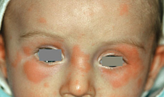
- Sudden onset of erythematous plaques of varied shape and size (Figure 1)
- Central pallor or red flare may be present
- Pruritus, burning
- Individual wheals appear, enlarge, and may coalesce
- Lesions are migratory and transient
- May appear anywhere on the body
- Associated angioedema is common
- Systemic features (flushing, shortness of breath, wheezing, headache, fatigue, fever, gastrointestinal distress, arthralgias, palpitations, lymphadenopathy, bruising)
- Systemic symptoms may be seen in the setting of blood transfusion induced serum sickness or a serum sickness-like reaction to a medication.
- Severe forms of allergic urticaria can lead to anaphylactic shock.
Clinical features of angioedema
- Non-pitting edema of the dermis and subcutaneous tissues
- Skin colored or slightly erythematous
- Often more painful than pruritic
- Mucous membrane involvement
- Gut edema (particularly in hereditary angioedema)
- Involvement of the pharynx or larynx can cause life-threatening asphyxiation
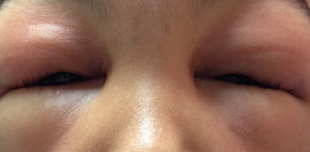
- Commonly affects the eyelids (Figure 2) and lips
Other diagnostic tests
- Skin prick allergy testing, fluoroimmunoassay or radioallergosorbent tests may be used in acute urticaria if a specific allergen is suspected.
- Although diagnostic testing for spontaneous chronic urticaria is not routine, autologous serum skin testing may be implemented to detect the circulating autoantibodies thought to induce disease. Disadvantages of this test include a high false positive rate, particularly in children with atopy, those with autoimmune thyroid disease, or patients taking certain common medications (i.e. antihistamines).
- Biopsy (if diagnosis is uncertain) is non-specific but may reveal edema and vasodilation in the dermis with an inflammatory infiltrate.
- C1 inhibitor assay can be performed if there is suspicion for hereditary angioedema.
Severity staging (used in chronic urticaria)
- The severity of chronic spontaneous urticaria is assessed based on the number of wheals present and the intensity of pruritis using the UAS7 scoring system.
- Quality of life scales including the Dermatology Life Quality Index have been validated for use in chronic urticaria.
Differential diagnosis
- Urticaria
- Contact dermatitis
- Facial cellulitis
- Papular urticaria (chronic hypersensitivity reaction to insect bites)
- Maculopapular cutaneous mastocytosis
- Erythema migrans
- Angioedema
- Contact dermatitis
- Facial cellulitis
- Facial lymphedema
- Superior vena cava syndrome
- Dermatomyositis heliotrope rash
- Blepharochalasis, Ascher syndrome
- Amyloidosis
Patient management: treatment and follow-up
As these conditions can be associated with laryngeal edema and anaphylaxis, airway stabilization and/or intramuscular epinephrine may be required. For any form of drug-induced angioedema (ie. aspirin/NSAID, ACE inhibitor) discontinuation of the offending agent is necessary.
- Medical therapy:
- Acute urticaria
- Single dose of a first-generation antihistamine (i.e. diphenhydramine), which acts on central H1 receptors causing significant sedation and anticholinergic side effects.
- Second generation antihistamines (i.e. fexofenadine, loratadine), which act mainly on peripheral H1 receptors, have fewer adverse effects and may be preferred if multiple doses are required.
- Corticosteroids (refractory disease)
- Chronic urticaria: treatment response is measured using the urticaria control test, in which patients score their symptoms and quality of life to determine if additional treatment is required.
- Second generation antihistamines
- Severe refractory disease:
- Omalizumab (anti-IgE monoclonal antibody)
- Cyclosporin (calcineurin inhibitor)
- Other less commonly used treatments (not approved by the Food and Drug Administration for chronic urticaria): methotrexate, tricyclic antidepressants, Anti-Tumor Necrosis Factor alpha agents, dapsone, phototherapy, plasmapheresis or intravenous immunoglobulin
- Mast cell mediated angioedema
- First or second-generation antihistamines.
- Oral glucocorticoids may reduce relapse rates.
- Immunosuppressive agents (i.e. cyclosporine) and omalizumab may be efficacious for chronic, refractory disease.
- Leukotriene antagonists and lipoxygenase inhibitors may be efficacious in NSAID induced disease.
- Bradykinin mediated angioedema
- Hereditary angioedema and acquired C1 inhibitor deficiency:
- First line: C1 inhibitor replacement.
- Second line: fresh frozen plasma (can rarely exacerbate disease), ecallantide (kallikrein inhibitor), icatibant (bradykinin receptor antagonist).
- Prophylaxis: tranexamic acid, lanadelumab (inhibitor of active kallikrein), anabolic steroids (increase circulating levels of C1 inhibitor), C1 inhibitor concentrate infusion.
- Rituximab has been used to successfully treat and induce long-lasting remission in acquired C1 inhibitor deficiency, whether or not there is an underlying lymphoproliferative disorder.
- ACE inhibitor induced angioedema
- Discontinuation of ACE Inhibitor.
- Surgical therapy: in rare cases, urticaria or angioedema can require an emergent surgical airway.
- Natural history and prognosis:
- Acute spontaneous urticaria
- Individual urticarial lesions typically resolve within 24 hours without scarring.
- Lesions may continue to develop and resolve for up to 6 weeks, after which disease is classified as chronic urticaria.
- Chronic spontaneous urticaria
- Lesions are slower to resolve than in the acute form, but typically change shape or reduce in size by 24 hours.
- After 5 years, 20% of patients continue to experience spontaneous whealing (Saini, 2018).
- Although extensive laboratory evaluation is not generally required, if there is clinical suspicion of an underlying autoimmune or infectious process, further workup may be initiated.
- Angioedema associated with urticaria
- Slower to resolve than urticaria, taking up to 72 hours.
- Hereditary angioedema
- Usually first presents in childhood, worsens during puberty, and persists into adulthood.
- Acquired C1 inhibitor deficiency
- Prognosis is complicated by the high likelihood that there is underlying lymphoproliferative or autoimmune disease. Those without concurrent disease at the time of diagnosis have been reported to subsequently develop lymphoproliferative disorders (Otani, 2017).
- Treatment of the provocative condition leads to resolution of angioedema in most cases.
- ACE inhibitor induced angioedema
- Angiotensin II receptor blockers (ARBs) are generally well-tolerated in these patients.
Complications
- Laryngeal edema with airway compromise
- Chronic urticaria can significantly impact quality of life.
- Those with disease refractory to antihistamines, which account for more than 50% of cases, have poorer outcomes on quality of life scales; specifically reporting issues with social relationships and adherence to medications (Kanamori, 2016).

Photograph courtesy of D@nderm Atlas of Clinical Dermatology.
Figure 1. Spontaneous acute urticaria of the bilateral periocular area in a child.

Photograph courtesy of Roman Shinder, MD.
Figure 2. Bilateral, isolated angioedema involving the upper and lower eyelids.
References and additional resources
- Zuberbier T. Urticaria. Allergy. 2003; 58(12): 1224-1234.
- Tsakok T & Flohr C. Pediatric urticaria. Immunology and Allergy Clinics of North America. 2014; 34(1): 117-139.
- Schaefer P. Acute and Chronic Urticaria: Evaluation and Treatment. Am Fam Physician. 2017 Jun 1;95(11):717-724.
- Kaplan A & Greaves M. Angioedema. Journal of American Academy of Dermatology. 2005; 53(3): 373-388.
- Weller K, Altrichter S, Ardelean E, et al. Chronische Urtikaria. Prävalenz, Verlauf, Prognosefaktoren und Folgen [Chronic urticaria. Prevalence, course, prognostic factors and impact]. Hautarzt. 2010;61(9):750‐757.
- Otani IM, Banerji A. Acquired C1 Inhibitor Deficiency. Immunol Allergy Clin North Am. 2017;37(3):497-511.
- Maurer M, Fluhr JW, Khan DA How to approach chronic inducible urticaria. J Allergy Clin Immunol: In Practice. 2018;6(4):1119-1130.
- Maurer M, Altrichter S, Bieber T, et al. Efficacy and safety of omalizumab in patients with chronic urticaria who exhibit IgE against thyroperoxidase. J Allergy Clin Immunol. 2011;128:202-209.
- Kolkhir P, Church M, Weller K, et al. Autoimmune chronic spontaneous urticaria: What we know and what we do not know. J Allergy Clin Immunol. 2017;139(6): 1772-1781.
- Radonjic-Hoesli S, Hofmeier KS, Micaletto S, et al. Urticaria and Angioedema: an Update on Classification and Pathogenesis. Clin Rev Allergy Immunol. 2018;54(1):88‐101.
- Zuberbier T. Urticaria. Allergy. 2003; 58(12): 1224-1234.
- Tsakok T & Flohr C. Pediatric urticaria. Immunology and Allergy Clinics of North America. 2014; 34(1): 117-139.
- Saini SS, Kaplan AP. Chronic Spontaneous Urticaria: The Devil’s Itch. J Allergy Clin Immunol Pract. 2018;6(4):1097‐1106.
- Kanamori KY, Alcantara CT, Motta AA, et al. Quality of life assessment in patients with chronic urticaria. J Allergy Clin Immunol. 2016;137(2).
